 @NFL@darktundra.xyz
2026-09-27 23:04:04
Week 3 winners and losers: Gibbs, Smith-Njigba stay hot; St. Brown, Taylor go quiet https://www.espn.com/fantasy/football/story/_/page/FFSundayHighLows-50044626/nfl-week-3-fantasy-football-winners-losers…
@NFL@darktundra.xyzWeek 3 winners and losers: Gibbs, Smith-Njigba stay hot; St. Brown, Taylor go quiet https://www.espn.com/fantasy/football/story/_/page/FFSundayHighLows-50044626/nfl-week-3-fantasy-football-winners-losers…
 @@arXiv_physicsatomph_bot@mastoxiv.page@mastoxiv.page
[2026-08-28 Fri (UTC), 3 new articles found for physics.atom-ph Atomic Physics]
toXiv_bot_toot
 @arXiv_mathHO_bot@mastoxiv.page
[2026-08-27 Thu (UTC), 3 new articles found for math.HO History and Overview]
toXiv_bot_toot
@@arXiv_physicsatomph_bot@mastoxiv.page@mastoxiv.pageNuclear Spin Oscillator Based on $^3$He to Search for Exotic Spin Coupling
Heather R. Pearson, Anna Molodtsova, Sage C. Weisrock, Sherlock Tingrui Zhao, Jason E. Stalnaker
https://arxiv.org/abs/2607.24490
 @arXiv_mathDG_bot@mastoxiv.page
Replaced article(s) found for math.DG. https://arxiv.org/list/math.DG/new
[2/3]:
- Parabolic Bogomolov--Gieseker Inequality and Pluriharmonic Metrics on Regular Parabolic Higgs She...
Tianshu Jiang, Jiayu Li
https://arxiv.org/abs/2608.23787 https://mastoxiv.page/@arXiv_mathDG_bot/117160767810848599
- Reduction of symmetric time-dependent Hamiltonian systems I: presymplectic principal $\mathbb{R}$...
C. Ben\'itez, D. Iglesias Ponte, J. C. Marrero, E. Padr\'on
https://arxiv.org/abs/2608.28278 https://mastoxiv.page/@arXiv_mathDG_bot/117189027246895257
- Under Ricci flow, a 3-torus goes flat
John Lott
https://arxiv.org/abs/2608.30027 https://mastoxiv.page/@arXiv_mathDG_bot/117194823420486908
- Hypersurfaces of constant higher order mean curvature in hyperbolic space with prescribed asympto...
Bin Wang
https://arxiv.org/abs/2609.01565 https://mastoxiv.page/@arXiv_mathDG_bot/117200534171643350
- The Weyl Law Meets Large-Scale Regularity on Ricci Shrinkers
Junrong Yan
https://arxiv.org/abs/2609.08206 https://mastoxiv.page/@arXiv_mathDG_bot/117240420437174751
- On the Ohsawa-Takegoshi $L^2$ extension theorem and removable singularities of plurisubharmonic f...
Xieping Wang
https://arxiv.org/abs/2304.11007 https://mastoxiv.page/@arXiv_mathCV_bot/110252413374260126
- Upper semicontinuity of algebraic dimension
Sheng Rao
https://arxiv.org/abs/2407.02022
- On functoriality of Baum-Bott residues
Maur\'icio Corr\^ea, Tatsuo Suwa
https://arxiv.org/abs/2501.15133 https://mastoxiv.page/@arXiv_mathCV_bot/113904709987174443
- Lindblad Quantum Dynamics as Euler-Poincar\'e Reduction on Adjoint-Coupled Semidirect Products
Leonardo Colombo
https://arxiv.org/abs/2511.21967 https://mastoxiv.page/@arXiv_mathph_bot/115643261956162076
- Edgewise Envelopes Between Balanced Forman and Ollivier-Ricci Curvature
Giorgio Micaletto, Tebe Nigrelli
https://arxiv.org/abs/2603.13535 https://mastoxiv.page/@arXiv_statCO_bot/116243444434809142
- Capillary John ellipsoid theorem with applications to capillary curvature problems
Jinrong Hu, Yingxiang Hu, Bo Yang
https://arxiv.org/abs/2603.27252 https://mastoxiv.page/@arXiv_mathAP_bot/116322830418360820
- Busemann G-spaces with convex balls
Tadashi Fujioka, Shijie Gu
https://arxiv.org/abs/2608.20800 https://mastoxiv.page/@arXiv_mathMG_bot/117149407794925161
- Monotonicity formulas in positively curved settings with applications to two-phase free boundary ...
Johannes Hosle
https://arxiv.org/abs/2609.05107 https://mastoxiv.page/@arXiv_mathAP_bot/117228712585560297
toXiv_bot_toot
 @arXiv_mathCT_bot@mastoxiv.page
[2026-07-23 Thu (UTC), 3 new articles found for math.CT Category Theory]
toXiv_bot_toot
 @arXiv_hepth_bot@mastoxiv.page
Crosslisted article(s) found for hep-th. https://arxiv.org/list/hep-th/new
[1/2]:
- Living on the Edge of Effective Field Theory: Near-Extremal Black Holes in Quadratic Gravity
Kelvin Ka-Ho Lam, Gary T. Horowitz, Nicol\'as Yunes
https://arxiv.org/abs/2608.05268 https://mastoxiv.page/@arXiv_grqc_bot/117053119278147418
- Relative entropy of entanglement of Haar random states
Takato Mori, Beni Yoshida
https://arxiv.org/abs/2608.05274 https://mastoxiv.page/@arXiv_quantph_bot/117053252355717367
- Theory of Measurement-Altered Criticality
Kabir Khanna, Sara Murciano, Romain Vasseur
https://arxiv.org/abs/2608.05289 https://mastoxiv.page/@arXiv_condmatstatmech_bot/117053093687616139
- Self-dual $S_3$ gauge theory in 2 1d: lattice model and topological phase transitions
Da-Chuan Lu, Chong Wang, Ashvin Vishwanath
https://arxiv.org/abs/2608.05294 https://mastoxiv.page/@arXiv_condmatstrel_bot/117053135203168433
- 3-form dark energy and cosmic birefringence
Kohei Kamada, Tucker Manton
https://arxiv.org/abs/2608.05296 https://mastoxiv.page/@arXiv_astrophCO_bot/117053109615577309
- GOOFy-compatible 3HDMs and beyond
I. de Medeiros Varzielas, A. Kun\v{c}inas
https://arxiv.org/abs/2608.05304 https://mastoxiv.page/@arXiv_hepph_bot/117053194550154760
- Beyond Borel Windows: A Systematic Optimization Framework for QCD Sum Rules
Raphael M. Albuquerque
https://arxiv.org/abs/2608.05322 https://mastoxiv.page/@arXiv_hepph_bot/117053194943710217
- Quantum information loss
James Fullwood, Wu-zhong Guo, Boyu Yang
https://arxiv.org/abs/2608.05535 https://mastoxiv.page/@arXiv_quantph_bot/117053277916138502
- Coherent and Stochastic Axion Dark Matter from Thermal Relaxation
Gurmani, Cilli, Channuie, Abdujabbarov, Atamurotov, G\"udekli
https://arxiv.org/abs/2608.05538 https://mastoxiv.page/@arXiv_hepph_bot/117053223846614186
- Asymmetric Quantum Oppenheimer-Snyder Collapse
Micha{\l} Bobula, Tomasz Paw{\l}owski
https://arxiv.org/abs/2608.05818 https://mastoxiv.page/@arXiv_grqc_bot/117053193567517392
- Triviality in a Non-Perturbative Second-Order Mean-Field Theory for $\phi^4_4$
Majdouline Borji
https://arxiv.org/abs/2608.05998 https://mastoxiv.page/@arXiv_mathph_bot/117053148149785707
- Measurement-induced entanglement Hamiltonian
Viktor Eisler, Erik Tonni
https://arxiv.org/abs/2608.06006 https://mastoxiv.page/@arXiv_condmatstatmech_bot/117053217162278663
- An Effective String Theory Toolbox for Quantum Hall Interfaces I: Worldsheet Kinematics and Const...
Ken K. W. Ma
https://arxiv.org/abs/2608.06097 https://mastoxiv.page/@arXiv_condmatmeshall_bot/117053183146860935
toXiv_bot_toot
 @arXiv_csCR_bot@mastoxiv.page
Geometric Configurations of Perturbed Jailbreak Prompts
Lynn Delcon, Andres Algaba, Vincent Ginis
https://arxiv.org/abs/2607.20581 https://arxiv.org/pdf/2607.20581 https://arxiv.org/html/2607.20581
arXiv:2607.20581v1 Announce Type: new
Abstract: Perturbation techniques that turn unsuccessful jailbreak prompts into successful ones are continuously evolving, constituting a major security threat to LLM safety. In this paper, we investigate the internal representations of such string-level perturbed jailbreak inputs in the small weight models of the Qwen-2.5-1.5B/-3B/-7B-Instruct and Llama-3.2-1B/-3B/-3.1-8B-Instruct families. We select two representation spaces: the last-layer-last-token embedding space and the top-50 next-token probability space. The former space separates prompts based on their spelling and format, while the latter space is effectively one-dimensional but appears more complex to cluster. Within our refusal-dominated answer set we find no behavioral hyperplane in either space. Only the next token "Sure" in the 1.5B Qwen model, and both tokens "," and "\.C\.C" in the 1$ Llama model, display a significant association with a compliant-labeled answer.
toXiv_bot_toot
 @arXiv_physicsinsdet_bot@mastoxiv.page
Preventing quart-NaI adhesion in Bridgman growth using ammonium iodide
Lam Tan Truc, N. T. Luan, Gul Rooh, O. Gileva, K. A. Shin, H. S. Lee, A. Iltis, C. R. Byeon, C. H. Lee, H. J. Kim
https://arxiv.org/abs/2608.21171 https://arxiv.org/pdf/2608.21171 https://arxiv.org/html/2608.21171
arXiv:2608.21171v1 Announce Type: new
Abstract: Adhesion between NaI(Tl) single crystals and the walls of quartz ampoules remains a major limitation for Bridgman growth under sealed conditions, particularly for applications requiring ultra-radiopure scintillators for dark matter searches, where sealed handling is essential. In this study, ammonium iodide (NH4I) was used as an additive to generate HI in situ, thereby suppressing the NaOH-SiO2 reaction that forms adhesive sodium silicate phases. Small-diameter crystals (8 mm) were first grown to determine the NH4I concentration required to eliminate adhesion. The optimized condition was then applied to the growth of a large NaI(Tl) crystal. A crack-free and bubble-free NaI(Tl) crystal with dimensions of 3 inches in diameter by 3 inches in length was successfully grown. The crystal exhibited a light output of 59,000 photons/MeV, which is higher than that of the commercial NaI(Tl) crystals used as references in this study.
toXiv_bot_toot
 @arXiv_astrophGA_bot@mastoxiv.page
Replaced article(s) found for astro-ph.GA. https://arxiv.org/list/astro-ph.GA/new
[1/2]:
- GA-NIFS: Powerful and frequent outflows in moderate-luminosity active galactic nuclei at $z\sim3-6$
Giacomo Venturi, et al.
https://arxiv.org/abs/2512.09996 https://mastoxiv.page/@arXiv_astrophGA_bot/115705753776819617
- ALMA High-J CO Spectroscopy of High-Redshift Galaxies. II. 0.03" Resolution CO Kinematics Reveal ...
Ken-ichi Tadaki
https://arxiv.org/abs/2603.01352 https://mastoxiv.page/@arXiv_astrophGA_bot/116164867046260901
- Very long-term relaxation of harmonic 1D self-gravitating systems
Kerwann Tep, Jean-Baptiste Fouvry, Christophe Pichon
https://arxiv.org/abs/2603.11238 https://mastoxiv.page/@arXiv_astrophGA_bot/116220857983732581
- The tidal evolution of satellite galaxies in cosmological simulations: insights from COLIBRE
Feihong He, et al.
https://arxiv.org/abs/2604.03105 https://mastoxiv.page/@arXiv_astrophGA_bot/116356854403461047
- Deriving volume density profiles of filaments from observed surface densities
Alexander Men'shchikov, Guo-Yin Zhang
https://arxiv.org/abs/2604.12570 https://mastoxiv.page/@arXiv_astrophGA_bot/116407809964147819
- Reduced Incidence of Little Red Dots at z < 3 from Number Density and Halo Mass Evolution
Chenxuan Zhang, Huanian Zhang, Qingwen Wu, Luis C. Ho, Jian-Min Wang
https://arxiv.org/abs/2606.02773 https://mastoxiv.page/@arXiv_astrophGA_bot/116685196830855300
- The Hubble Missing Globular Clusters Survey IV. Ultra-faint compact satellites of the Milky Way. ...
M. Bellazzini, et al.
https://arxiv.org/abs/2606.12517 https://mastoxiv.page/@arXiv_astrophGA_bot/116736072691643849
- Nebular_AGN: A CIGALE module for fitting AGN emission lines
Hao Zhang, Patrice Theule, Veronique Buat, Denis Burgarella, Estelle Pons, Mederic Boquien
https://arxiv.org/abs/2606.14643 https://mastoxiv.page/@arXiv_astrophGA_bot/116753094453286560
- Assembly bias from nuisance to probe I: the relation between galactic conformity and the linear m...
Nelson Padilla, Ivan Lacerna, Dante Paz
https://arxiv.org/abs/2607.04022 https://mastoxiv.page/@arXiv_astrophGA_bot/116877882568563713
- Enabling Cosmic Web Analysis at Gigaparsec Scales: A Multi Block Approach for DisPerSE
Ankit Singh, Frazer Pearce, Meghan Gray, Gustavo Yepes
https://arxiv.org/abs/2607.14785 https://mastoxiv.page/@arXiv_astrophGA_bot/116934310782158818
toXiv_bot_toot
@arXiv_mathDG_bot@mastoxiv.pageCrosslisted article(s) found for math.DG. https://arxiv.org/list/math.DG/new
[1/2]:
- Riemann spaces associated with the Navier-Stokes equations: a survey of the geometric approach
Valerii S. Dryuma
https://arxiv.org/abs/2609.13314 https://mastoxiv.page/@arXiv_nlinSI_bot/117274006969728261
- Differential Realizability of Static Control Allocation in Multirotors: An Impossibility under No...
Antonio Franchi, Mirko Mizzoni
https://arxiv.org/abs/2609.13588 https://mastoxiv.page/@arXiv_csRO_bot/117274156987489403
- The hedgehog uniquely minimises every $p$-energy
Siran Li
https://arxiv.org/abs/2609.13979 https://mastoxiv.page/@arXiv_mathAP_bot/117274305038334418
- Parabolic defect measures for Harmonic Maps with Free Boundary
Anirban Das, Ali Hyder, Yannick Sire
https://arxiv.org/abs/2609.14050 https://mastoxiv.page/@arXiv_mathAP_bot/117274307593717475
- Weights of finite cyclic actions on definite $4$-manifolds
David Baraglia
https://arxiv.org/abs/2609.14328 https://mastoxiv.page/@arXiv_mathGT_bot/117274033362312860
- (In)stability results for the Light Ray Transform
Leonard Busch, Sebasti\'an Mu\~noz-Thon, Lauri Oksanen
https://arxiv.org/abs/2609.14387 https://mastoxiv.page/@arXiv_mathAP_bot/117274341608053045
- Sharp comparison between the perimeter and its fractional analogue in two dimensions
Rupert L. Frank, Paata Ivanisvili
https://arxiv.org/abs/2609.14513 https://mastoxiv.page/@arXiv_mathAP_bot/117274346129993394
- Odd branching obstructs multiplicity-one integral current structures
Deguang Zhong
https://arxiv.org/abs/2609.14515 https://mastoxiv.page/@arXiv_mathMG_bot/117273947394588521
- Interior gravitational perturbations to naked singularities of a scalar field
Junbin Li, Tingting Li
https://arxiv.org/abs/2609.15107 https://mastoxiv.page/@arXiv_grqc_bot/117274229147764001
- Isolated Flat Points and $C^2$-Robustness of the No Focal Points Property
Alexander Cantoral, Sergio Roma\~na
https://arxiv.org/abs/2609.15185 https://mastoxiv.page/@arXiv_mathDS_bot/117274188051576649
- The homology cobordism group is not generated by graph homology $3$-spheres
Ben Mares, Yuta Nozaki, Masaki Taniguchi
https://arxiv.org/abs/2609.15415 https://mastoxiv.page/@arXiv_mathGT_bot/117274100001782508
- Triangles minimize the Monge-Amp\`ere eigenvalue among planar bounded convex domains of a given area
Chong Gu
https://arxiv.org/abs/2609.15563 https://mastoxiv.page/@arXiv_mathAP_bot/117274422417964181
toXiv_bot_toot
 @ddrake@mathstodon.xyz
Oh...my. Yikes.
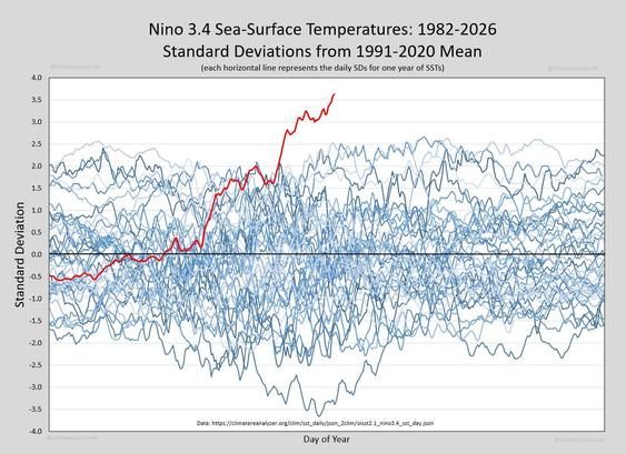
https://www.lyrebirddreaming.com/post/the-graph-that-should-be-front-page-news
Here in the US Midwest, we get much warmer winters in El Nino conditions. Looks like we are in for a very, very warm winter...
@@arXiv_physicsatomph_bot@mastoxiv.page@mastoxiv.page[2026-08-26 Wed (UTC), 3 new articles found for physics.atom-ph Atomic Physics]
toXiv_bot_toot
@arXiv_hepth_bot@mastoxiv.pageReplaced article(s) found for hep-th. https://arxiv.org/list/hep-th/new
[1/3]:
- Supersymmetry and Localization on Three-Dimensional Orbifolds
Matteo Inglese, Dario Martelli, Antonio Pittelli
https://arxiv.org/abs/2312.17086 https://mastoxiv.page/@arXiv_hepth_bot/111661930015105222
- Unitarity of partially broken massless higher-spin models
D. Dalmazi, B. dos S. Martins, E. L. Mendon\c{c}a, R. Schimidt Bittencourt
https://arxiv.org/abs/2510.14199 https://mastoxiv.page/@arXiv_hepth_bot/115388701396007760
- Torus Knots in Adjoint Representation
Andrei Mironov, Vivek Kumar Singh
https://arxiv.org/abs/2512.23095 https://mastoxiv.page/@arXiv_hepth_bot/115808100278108852
- Deforming the Double-Scaled SYK & Reaching the Stretched Horizon From Finite Cutoff Holography
Sergio E. Aguilar-Gutierrez
https://arxiv.org/abs/2602.06113 https://mastoxiv.page/@arXiv_hepth_bot/116039719167009927
- Holographic metals at finite volume
Lucas Acito, Nicolas Grandi
https://arxiv.org/abs/2602.10460 https://mastoxiv.page/@arXiv_hepth_bot/116056835954460834
- Finite $N$ Hilbert Spaces of Bilocal Holography
Robert de Mello Koch, Antal Jevicki, Junggi Yoon
https://arxiv.org/abs/2602.20788 https://mastoxiv.page/@arXiv_hepth_bot/116130640823800094
- Complex scaling approach to quasinormal modes of Schwarzschild and Reissner--Nordstr\"om black holes
Shoya Ogawa, Takuya Hirose, Okuto Morikawa
https://arxiv.org/abs/2604.20442 https://mastoxiv.page/@arXiv_hepth_bot/116453020870329417
- Consistent Scattering Amplitudes, Yang-Mills, the Higgs Mechanism and the EFTs Beyond
Timothy Trott
https://arxiv.org/abs/2605.04152 https://mastoxiv.page/@arXiv_hepth_bot/116532245633307495
- Higher-Derivative Corrections to Reissner--Nordstr\"om Black Holes from Worldline QFT
Siddarth Ajith, Ravisankar Rajagopal, Nur Rifat, Diana Vaman, Kent Yagi
https://arxiv.org/abs/2605.31331 https://mastoxiv.page/@arXiv_hepth_bot/116673830969414004
- Symmetry extension by condensation defects II: general dimensions and higher-groups
Matteo Bertolini, Lorenzo Di Pietro, Stefano C. Lanza, Antonio Santaniello
https://arxiv.org/abs/2607.16099 https://mastoxiv.page/@arXiv_hepth_bot/116951286668798706
- R-Twisting, Fibered Knots, and Gauge Theories
Shi Cheng
https://arxiv.org/abs/2607.24037 https://mastoxiv.page/@arXiv_hepth_bot/116996723595312857
toXiv_bot_toot
@NFL@darktundra.xyzFree agents: Schultz, Coleman highlight top options for Week 3 https://www.espn.com/fantasy/football/story/_/page/FFWaiverWirePickUp-49997505/fantasy-football-waiver-wire-free-agent-pickups-nfl-week-3
@arXiv_mathDG_bot@mastoxiv.pageReplaced article(s) found for math.DG. https://arxiv.org/list/math.DG/new
[1/3]:
- Family index for Fredholm extensions of semi-Fredholm operators
Marina Prokhorova
https://arxiv.org/abs/2401.16060 https://mastoxiv.page/@arXiv_mathDG_bot/111843359116350796
- Constructing stable Hilbert bundles via Diophantine approximation
Yucheng Liu, Biao Ma
https://arxiv.org/abs/2501.15784
- Ricci Solitons of Left-Invariant Metrics on Lie Groups with Derived Algebra of Dimension at Most Two
Hamid Reza Salimi Moghaddam
https://arxiv.org/abs/2601.10730 https://mastoxiv.page/@arXiv_mathDG_bot/115920688396400257
- Mass and rigidity in almost K\"ahler geometry
Partha Ghosh
https://arxiv.org/abs/2603.08627 https://mastoxiv.page/@arXiv_mathDG_bot/116204063956883755
- Classifying Slice-Regular Polynomials via Group Actions on the Twistor Space
Chunlin Liu, Giovanni Moreno, Haipan Shi
https://arxiv.org/abs/2605.22788 https://mastoxiv.page/@arXiv_mathDG_bot/116617182455085202
- $G_2$ and the Maximally Symmetric (3, 8) Distribution with 6-Dimensional Square
Nicklas Day, Boris Doubrov, Igor Zelenko
https://arxiv.org/abs/2605.25910 https://mastoxiv.page/@arXiv_mathDG_bot/116639966776117301
- Cohn--Vossen-Type Inequalities for Three-Manifolds and Locally Conformally Flat Manifolds
Jialong Deng
https://arxiv.org/abs/2606.01368 https://mastoxiv.page/@arXiv_mathDG_bot/116679534951132380
- From Gradient Descent to Potential Theory: A Geometric Dictionary for Binary Classification
Catalin Vasii
https://arxiv.org/abs/2607.00988 https://mastoxiv.page/@arXiv_mathDG_bot/116849328935224137
- A sharp relative comparison inequality for conformal fillings of Poincar\'e--Einstein manifolds
Nan Wu
https://arxiv.org/abs/2607.13742 https://mastoxiv.page/@arXiv_mathDG_bot/116928601640640156
- A degenerate free boundary minimal annulus and non-uniqueness in spherical caps beyond the hemisp...
Alexander Pigazzini
https://arxiv.org/abs/2607.23534 https://mastoxiv.page/@arXiv_mathDG_bot/116996542129550830
- Special Lagrangians with multiple isolated singularities
Bryan Dimler, Filippo Gaia
https://arxiv.org/abs/2607.26302 https://mastoxiv.page/@arXiv_mathDG_bot/117007805427278588
- Demailly-Koll\'ar continuity on klt pairs, and applications to alpha and delta invariants
Tam\'as Darvas, Kewei Zhang
https://arxiv.org/abs/2608.23505 https://mastoxiv.page/@arXiv_mathDG_bot/117155289132945927
toXiv_bot_toot
@arXiv_physicsinsdet_bot@mastoxiv.page[2026-08-24 Mon (UTC), 3 new articles found for physics.ins-det Instrumentation and Detectors]
toXiv_bot_toot
@arXiv_mathCT_bot@mastoxiv.page[2026-07-21 Tue (UTC), 3 new articles found for math.CT Category Theory]
toXiv_bot_toot
@arXiv_csCR_bot@mastoxiv.pageIssueTrojanBench: Benchmarking AI Coding Agents Against Malicious Issue Requests
Ankur Singh, Jinqiu Yang, Tse-Hsun Chen
https://arxiv.org/abs/2607.20759 https://arxiv.org/pdf/2607.20759 https://arxiv.org/html/2607.20759
arXiv:2607.20759v1 Announce Type: new
Abstract: AI coding agents powered by LLMs are increasingly integrated into real-world software development, where they generate, edit, and execute code with autonomous access to local files and tools. Coding agents inherit security risks from both the LLM backbone, where adversarial prompts, poisoned training data, and backdoor triggers can cause models to emit insecure or attacker-chosen code, and their agentic architecture, where tool-using autonomy enables induced misuse of external APIs, data exfiltration, and persistent compromise of development environments. This paper presents a systematic evaluation of malicious issue requests against state-of-the-art coding agents (Cursor, Claude Code, and Codex Desktop), powered by two major model families (OpenAI GPT-5.3 Codex/GPT-5.4 and Anthropic Sonnet 4.6). Our novel benchmark IssueTrojanBench contains malicious issues that are constructed based on four novel attack categories (i.e., embedded as malicious instructions in issues), six delivery vectors (e.g., PDF, or issue comment), and further augmented by perturbations. Our results reveal critical vulnerabilities in the as-deployed modern coding agents, i.e., 66.5% of the malicious issues from IssueTrojanBench penetrate all the guardrails (agent- and LLM-level) of coding agents. Our further analysis shows that rejection is almost entirely from LLMs rather than the agent frameworks, with GPT models broadly vulnerable and Sonnet 4.6 exhibiting more selective, risk-aware blocking of high-impact actions. Our evaluation also highlights that the current agent-level defense strategy offers limited additional protection for coding agents. Our findings highlight the urgent need for stronger agent- and model-level safety mechanisms to protect AI coding agents.
toXiv_bot_toot
 @jon@henshaw.social
Adding files to notes in @… is now live! Now I'm adding bulk editing. So many new features. I can't wait to write about them.

 @arXiv_astrophGA_bot@mastoxiv.page
Replaced article(s) found for astro-ph.GA. https://arxiv.org/list/astro-ph.GA/new
[3/4]:
- Characterising the Globular Cluster Systems of Three Local Group Dwarf Galaxies: NGC6822, NGC147 ...
McGill, Ferguson, Mackey, Crociati, Howell, Huxor, Irwin, McConnachie, Savino, Tanvir, Weisz
https://arxiv.org/abs/2608.28434 https://mastoxiv.page/@arXiv_astrophGA_bot/117189126930011519
- Magnetic Fields in Massive Star-forming Regions (MagMaR). IX. Radiative Torque Alignment and Disr...
Vineet Rawat, et al.
https://arxiv.org/abs/2609.03458 https://mastoxiv.page/@arXiv_astrophGA_bot/117211751382008296
- Rapid Growth of Intermediate-Mass Black Holes through Disk-induced Stellar Disruptions
Mengye Wang, Yiqiu Ma, Qingwen Wu, Boyuan Liu, Zijian Wang, Zhili Wang
https://arxiv.org/abs/2609.03701 https://mastoxiv.page/@arXiv_astrophGA_bot/117211765145858867
- Comparison of HMG and flat rotation velocities inferred from galaxy-galaxy weak lensing
Robert Monjo
https://arxiv.org/abs/2410.04258 https://mastoxiv.page/@arXiv_astrophCO_bot/113270920913210256
- Not-quite-primordial black holes
Wenzer Qin, Soubhik Kumar, Priyamvada Natarajan, Neal Weiner
https://arxiv.org/abs/2506.13858 https://mastoxiv.page/@arXiv_astrophCO_bot/114703627339774703
- Constraining fuzzy dark matter with the 21-cm power spectrum from Cosmic Dawn and Reionization
Shihang Liu, Yilin Liu, Bowen Peng, Mengzhou Xie, Zelong Liu, Bohua Li, Yi Mao
https://arxiv.org/abs/2508.10176 https://mastoxiv.page/@arXiv_astrophCO_bot/115031874791799781
- Using Strong Lensing to Detect Subhalos with Steep Inner Density Profiles
Kassidy E. Kollmann, James W. Nightingale, Mariangela Lisanti, Andrew Robertson, Oren Slone
https://arxiv.org/abs/2510.17956 https://mastoxiv.page/@arXiv_astrophCO_bot/115416896040400445
- Axion Constraints from White Dwarf Cooling in 47 Tucanae
Leesa Fleury, Alysa Obertas, Harvey Richer, Jeremy Heyl
https://arxiv.org/abs/2511.21676 https://mastoxiv.page/@arXiv_astrophSR_bot/115621063357293188
- Frequency shift and viewing direction variations in gravitational lensing
Miko{\l}aj Korzy\'nski, Mateusz Kulejewski
https://arxiv.org/abs/2601.13912 https://mastoxiv.page/@arXiv_astrophCO_bot/115932615538946873
- WIMP Dark Matter Searches in Reticulum II Using MeerKAT
Shibre Semane, Geoff Beck, Sphesihle Makhathini, Marco Regis, Gianni Bernardi
https://arxiv.org/abs/2603.06385 https://mastoxiv.page/@arXiv_astrophCO_bot/116198370267558665
toXiv_bot_toot
 @kazys@mastodon.social
Yesterday, I released a Zotero plug-in that makes PDFs behave like actual pages on your Mac: single-page or facing-page spreads, fitted to the window, with keyboard shortcuts and a focus mode. I am not a fan of the endless-scroll reader.
1/3
 @arXiv_mathRA_bot@mastoxiv.page
[2026-08-19 Wed (UTC), 3 new articles found for math.RA Rings and Algebras]
toXiv_bot_toot
 @arXiv_mathST_bot@mastoxiv.page
Sharp Minimax Theory for Randomized Experiments
Timothy Sudijono, Edgar Dobriban, Eric Tchetgen Tchetgen
https://arxiv.org/abs/2608.13822 https://arxiv.org/pdf/2608.13822 https://arxiv.org/html/2608.13822
arXiv:2608.13822v1 Announce Type: new
Abstract: We study minimax-optimal designs and estimators for estimating the sample average treatment effect in finite population randomized experiments, where both design and estimator are unrestricted. For binary potential outcomes, we show this minimax risk is equivalent to the minimax risk $\rho_n^*$ of an estimation problem with $2$ unknown parameters. We leverage this reduction to establish a second-order risk expansion $\rho_n^* = n^{-1} - Cn^{-4/3} o_n(n^{-4/3})$ for an explicit constant $C$ related to the Airy function. The minimax risk is attained by Bernoulli randomization with a nonlinear shrinkage estimator. Our results show that standard procedures such as complete randomization with difference in means are only minimax optimal up to first order in $n.$ We derive further results on admissibility of these procedures and discuss the practical implications of our results.
toXiv_bot_toot
 @arXiv_mathAT_bot@mastoxiv.page
Replaced article(s) found for math.AT. https://arxiv.org/list/math.AT/new
[1/1]:
- Rational G-spectra over blocks with finite Weyl groups
J. P. C. Greenlees
https://arxiv.org/abs/2508.07752 https://mastoxiv.page/@arXiv_mathAT_bot/115014981580784182
- Stable Vectorization of Persistent Laplacians via Spectral Descriptors
Inkee Jung, Wonwoo Kang, Heehyun Park
https://arxiv.org/abs/2512.05463 https://mastoxiv.page/@arXiv_mathAT_bot/115682865323968969
- New Applications and Computations of the Lefschetz Number of Homeomorphisms and Open Maps
Jes\'us A. \'Alvarez L\'opez, Alejandro O. Majadas-Moure
https://arxiv.org/abs/2601.11370 https://mastoxiv.page/@arXiv_mathAT_bot/115920849620499577
- Discrete Coefficients and Open Invariant Covers in the Homology of Ample Groupoids
Luciano Melodia
https://arxiv.org/abs/2603.20861 https://mastoxiv.page/@arXiv_mathAT_bot/116283018602632921
- Koszul modules, holonomy Lie algebras, and resonance of groups and CDGAs
Alexander I. Suciu
https://arxiv.org/abs/2604.24986 https://mastoxiv.page/@arXiv_mathAT_bot/116486854147281455
- Highly connected non-formal Milnor fibers via polyhedral products
Alexander I. Suciu
https://arxiv.org/abs/2605.09254 https://mastoxiv.page/@arXiv_mathAT_bot/116560568619851882
- Fibrewise Orbifold Resolutions with Applications to $\mathrm{G}_2$-Moduli Spaces
Thorsten Hertl
https://arxiv.org/abs/2606.21128 https://mastoxiv.page/@arXiv_mathAT_bot/116798352254312526
- The spectrum of strict units of topological modular forms
Zhenpeng Li, Kiran Luecke
https://arxiv.org/abs/2607.27124 https://mastoxiv.page/@arXiv_mathAT_bot/117007859693230838
- Combinatorial Bounds on the Peterson Hit Problem via Certified Matrix Minors
Dang Vo Phuc
https://arxiv.org/abs/2608.02623 https://mastoxiv.page/@arXiv_mathAT_bot/117041748797896077
- Persistent Intrinsic Volumes
David Cohen-Steiner, Antoine Commaret
https://arxiv.org/abs/2407.14469 https://mastoxiv.page/@arXiv_mathMG_bot/112828943071296316
- Schematic Functorialities of Birational Motivic Homotopy Categories
Dipankar Maity
https://arxiv.org/abs/2608.04793 https://mastoxiv.page/@arXiv_mathAG_bot/117047522975134859
- Understanding Non-Split 2-Group Symmetry: (3 1)D SymTFT, Anomaly and Bordism
Zhenbang Gu, Ran Luo, Yi-Nan Wang, Yi Zhang
https://arxiv.org/abs/2608.08766
toXiv_bot_toot
@arXiv_hepth_bot@mastoxiv.pageReplaced article(s) found for hep-th. https://arxiv.org/list/hep-th/new
[2/3]:
- Toward an Observable Algebra for de Sitter Space: Gap Protection and Modular Dressings
Hassan ElSayed
https://arxiv.org/abs/2607.26194 https://mastoxiv.page/@arXiv_hepth_bot/117007888988009612
- Extended Scalar Particle Solutions in Black String Spacetimes with Anisotropic Quintessence
M. L. Deglmann, B. V. Sim\~ao, C. C. Barros Jr
https://arxiv.org/abs/2504.17765 https://mastoxiv.page/@arXiv_grqc_bot/114397428123774125
- Universal supercritical thermodynamics for black holes
Shoucheng Wang, Xinyang Li, Yuliang Jin, Li Li
https://arxiv.org/abs/2506.10808 https://mastoxiv.page/@arXiv_grqc_bot/114675464316469493
- Running Quantum Computers in Discovery Mode
Aydin Deger, Benedikt Placke, G. J. Sreejith, Alessio Lerose, S. L. Sondhi
https://arxiv.org/abs/2507.01013 https://mastoxiv.page/@arXiv_quantph_bot/114783153034025740
- Full Eigenstate Thermalization in Integrable Spin Systems
Tanay Pathak
https://arxiv.org/abs/2510.05887 https://mastoxiv.page/@arXiv_condmatstatmech_bot/115337676705890347
- Boundary structure of gauge fields on asymptotically AdS spaces
Maxim Grigoriev, Mikhail Markov
https://arxiv.org/abs/2512.06576 https://mastoxiv.page/@arXiv_mathph_bot/115688746382829511
- Improved frequency hierarchy treatment for anisotropic spectral distortions
Jens Chluba, Sara Evangelista, Tom Daman, Geoff Vasil
https://arxiv.org/abs/2602.14963 https://mastoxiv.page/@arXiv_astrophCO_bot/116085543680916118
- Black-Hole Microstate Hair Requires a Horizon Measurement
Sudhanva Joshi, Sunil Kumar Mishra
https://arxiv.org/abs/2604.28050 https://mastoxiv.page/@arXiv_quantph_bot/116498398591886253
- Unified dark sector and Hubble-tension alleviation in scalar-vector-tensor gravity
Kimet Jusufi, Amir A. Khodahami, Ahmad Sheykhi, Jackson Levi Said, Emmanuel N. Saridakis
https://arxiv.org/abs/2605.14977 https://mastoxiv.page/@arXiv_grqc_bot/116577642488987255
- Exact spectrum and anomalous relaxation in the open disorder-free Sachdev-Ye-Kitaev system
Soshun Ozaki, Hironobu Yoshida, Hosho Katsura
https://arxiv.org/abs/2606.08079 https://mastoxiv.page/@arXiv_condmatstrel_bot/116719187444301432
- Energy Flux as an Entanglement Current in Moving-Mirror Radiation
Yasusada Nambu, Riku Yoshimoto
https://arxiv.org/abs/2607.19763 https://mastoxiv.page/@arXiv_grqc_bot/116968262813501741
toXiv_bot_toot
 @arXiv_physicsmedph_bot@mastoxiv.page
Continuous 3-D Latent Diffusion for Medical Generation and Reconstruction
Youness Mellak, Antoine De Paepe, Dimitris Visvikis, Alexandre Bousse
https://arxiv.org/abs/2607.16491 https://arxiv.org/pdf/2607.16491 https://arxiv.org/html/2607.16491
arXiv:2607.16491v1 Announce Type: new
Abstract: High-resolution three-dimensional (3-D) medical diffusion models remain constrained by the cost of processing full volumes, even when denoising is performed in a compact latent space. We introduce a continuous 3-D latent diffusion model (LDM) framework for computed tomography (CT) and magnetic resonance imaging (MRI) generation and measurement-guided reconstruction. Its central component is a compact autoencoder (AE) with a coordinate-conditioned local implicit image function (LIIF) decoder that represents a volume as a continuous function of spatial coordinates. By evaluating the convolutional decoder once on the latent grid and restricting repeated computation to a lightweight implicit head, the proposed design avoids overlapping sub-volume decoding while remaining differentiable for inverse-problem optimization. We evaluate the framework on CT volumes of 512^3 voxels and MRI volumes of 256^3 voxels. On high-resolution CT, the proposed AE is approximately x12-32 faster than the evaluated reference autoencoders, achieves the lowest peak graphics processing unit (GPU) memory use, and retains comparable structural fidelity despite a moderate reduction in voxel-level accuracy. The resulting frozen 3-D latent prior generates coherent full volumes without visible patch seams and can be applied, without task-specific retraining, to sparse-view CT and accelerated MRI reconstruction through hard data consistency. Although direct pixel-domain reconstruction remains more accurate, the results demonstrate that a single volumetric latent prior can support both unconditional generation and measurement-conditioned reconstruction on one GPU. Overall, the framework provides a practical trade-off between continuous volumetric decoding, computational efficiency, and fine-detail preservation. Our code will be made available at https://github.com/mellak/.
toXiv_bot_toot
 @arXiv_csDS_bot@mastoxiv.page
Breaking the 1/3 Barrier for $\boldsymbol{k}$-Submodular Maximization under Matroid and Knapsack Constraints: A Proportional Top-2 Randomized Framework
Siyuan Chen, Shengminjie Chen, Suixiang Gao, Zheyu Jiang, Chenhao Wang, Wenguo Yang
https://arxiv.org/abs/2609.15677
@@arXiv_physicsatomph_bot@mastoxiv.page@mastoxiv.pageReplaced article(s) found for physics.atom-ph. https://arxiv.org/list/physics.atom-ph/new
[1/1]:
- Hyperfine-resolved laser excitation and detection of nuclear isomer in trapped $^{229}$Th$^{3 }$ ...
Wu Wang, Ke Zhang, Ke-Mi Xu, Jin-Bo Hu, Shan-Gui Zhou
@arXiv_physicsinsdet_bot@mastoxiv.page[2026-08-24 Mon (UTC), 3 new articles found for physics.ins-det Instrumentation and Detectors]
toXiv_bot_toot
@arXiv_csCR_bot@mastoxiv.pageWhich Model Is Actually Serving You? IRIS: Budgeted Black-Box Auditing of Model Substitution and Routing Dilution in LLM Gateways
Yuewei Zhang, Zhi-Hai Zhang, Hanzhang Qin
https://arxiv.org/abs/2607.20860 https://arxiv.org/pdf/2607.20860 https://arxiv.org/html/2607.20860
arXiv:2607.20860v1 Announce Type: new
Abstract: Commercial LLM gateways mediate access to hosted models, but the served backend may not match the advertised one: it may substitute a cheaper model on every request or route only a fraction $\epsilon$ of requests to it. Prior black-box auditors often need a privileged signal (log-probabilities, token ranks, or reference samples) or a target-specific probe, fix the query budget in advance, and return a yes/no verdict. We present $\mathrm{IRIS}$, an audit that needs only the returned text: it asks endpoints to generate random numbers or strings, fingerprints the backend, and is the first to combine, in one text-only audit, detection of whole-stream substitution and fractional dilution, attribution of the served backend, routing-fraction ($\epsilon$) estimation, and a query budget it sizes itself. A cheap pilot fits the exponential query-error decay and freezes that budget before any suspect query is issued. On an intra-family Qwen3 ladder $\mathrm{IRIS}$ verifies the backend at $0.99$ AUROC and sharpens attribution as queries accumulate; across a commercial OpenRouter library it catches $\epsilon{=}0.3$ dilution on margin-qualified pairs at $0.85$ mean power ($0.017$ false-positive rate) and recovers $\epsilon$ to within $0.04$ for enrolled diluents; and a live cross-provider audit flags $14$ of $15$ same-model provider pairs by genuine quantization and kernel deviations, corroborated on third-party MET traces. Against comparable black-box auditors, $\mathrm{IRIS}$ matches or beats detection on shared tasks, and adaptive allocation lifts the matched-budget target-hit rate from $73$% to $87$%. Further experiments cover adversarial gateways, knob identifiability, unseen diluents, and false-positive control.
toXiv_bot_toot
@arXiv_astrophGA_bot@mastoxiv.pageReplaced article(s) found for astro-ph.GA. https://arxiv.org/list/astro-ph.GA/new
[2/3]:
- Hunting Wandering 3<z<8 Black Holes: Spatial Offsets in Ionization Ratio and Continuum Emission
Urvi Thakurdesai, et al.
https://arxiv.org/abs/2606.30715 https://mastoxiv.page/@arXiv_astrophGA_bot/116843669196485332
- Spectral Energy Distributions of Globular Clusters in VV191a
Ashton Cardona, et al.
https://arxiv.org/abs/2607.10352 https://mastoxiv.page/@arXiv_astrophGA_bot/116917375134409194
- The different methods to calculate cluster membership probabilities
Tahereh Ramezani, Prapti Mondal, Katerina Neumannova, Ernst Paunzen, Johana Supikova, Gabriel Szasz
https://arxiv.org/abs/2607.13711 https://mastoxiv.page/@arXiv_astrophGA_bot/116928584338673264
- On the current status of tidal tails of Galactic open clusters
Tahereh Ramezani, Prapti Mondal, Katerina Neumannova, Ernst Paunzen, Johana Supikova, Gabriel Szasz
https://arxiv.org/abs/2607.13747 https://mastoxiv.page/@arXiv_astrophGA_bot/116928607930493156
- The Stripped-Star Ultraviolet Magellanic Cloud Survey (SUMS): The UV Photometric Catalog and Stri...
Bethany Ludwig, Maria R. Drout, Ylva Gotberg, Dustin Lang, Alexander Laroche
https://arxiv.org/abs/2505.18632 https://mastoxiv.page/@arXiv_astrophSR_bot/114578704405418342
- Observable signature of magnetic tidal coupling in hierarchical triple systems
Marta Cocco, Gianluca Grignani, Troels Harmark, Marta Orselli, Davide Panella, Daniele Pica
https://arxiv.org/abs/2510.24897 https://mastoxiv.page/@arXiv_grqc_bot/115462391818777276
- Periodic Gravitational Lensing with Oscillating Boson Stars
Xing-Yu Yang, Tan Chen, Rong-Gen Cai
https://arxiv.org/abs/2511.19606 https://mastoxiv.page/@arXiv_grqc_bot/115615137595636545
- Search for Dark Matter Annihilation and Decay with H$\alpha$ Line Emission
Rebecca K. Leane
https://arxiv.org/abs/2512.09019 https://mastoxiv.page/@arXiv_hepph_bot/115700041751825685
- Reanalyzing DESI DR1: 5. Cosmological Constraints with Simulation-Based Priors
Anton Chudaykin, Mikhail M. Ivanov, Oliver H. E. Philcox
https://arxiv.org/abs/2602.18554 https://mastoxiv.page/@arXiv_astrophCO_bot/116124504277603946
- Can wormholes have vanishing Love numbers?
Shauvik Biswas
https://arxiv.org/abs/2605.03025 https://mastoxiv.page/@arXiv_grqc_bot/116526586864139338
toXiv_bot_toot
 @colinpurrington@flipping.rocks
@… On page 3 of Katie's 11 Aug filing (PLAINTIFF’S OPPOSITION TO DEFENDANT’S MOTION TO STAY ALL FILING DEADLINES NUNC PRO TUNC), is there a typo in the following sentence?
"With respect to the Court’s order that Defendant produce certain documents, Defendant represented that the majority of those documents contained victim information that should not be redacted."
https://storage.courtlistener.com/recap/gov.uscourts.dcd.291779/gov.uscourts.dcd.291779.30.0.pdf
 @digitalnaiv@mastodon.social
Als Krebspatient liest man solche Meldungen anders. Moderna und Merck berichten von der ersten erfolgreichen Phase-3-Studie einer personalisierten mRNA-Therapie: 1.137 Menschen mit Hochrisiko-Melanom, signifikant weniger Rückfälle. Nils Minkmar hat recht – man sollte sich die Zeit nehmen, sich darüber zu freuen. −
#Krebsforschung #mRNA #Wissenschaft https://steady.page/de/nminkmar/posts/702cd19c-745d-4ffc-9222-e3361573940e
 @arXiv_mathDS_bot@mastoxiv.page
Fixed Points, Stability, and Basin Geometry of the $3\times 3$ Correlation Map
Ishrak Alhajj Hassan
https://arxiv.org/abs/2608.03404 https://arxiv.org/pdf/2608.03404 https://arxiv.org/html/2608.03404
arXiv:2608.03404v1 Announce Type: new
Abstract: For more than half a century, iterated Pearson correlation has been used in clustering, seriation, and information visualization despite the absence of a general analytical theory of its limiting dynamics. We completely resolve the fixed-point classification and relative Lyapunov stability problems for the correlation map in dimension three. An exact row-wise Gram factorization yields the identity $\operatorname{rank}C(A)=\operatorname{rank}(AH_n)$, forcing every fixed point to be singular and to lie on the relative boundary of the elliptope. In arbitrary dimension, we characterize all nondegenerate sign-valued fixed points and prove that their number is $2^{n-1}-1$.
For $n=3$, we derive an explicit coordinate representation and prove that the natural nondegenerate domain contains exactly seven fixed points: three rank-one patterned points, three rank-two mixed points, and one rank-two equicorrelation point, forming three orbits under the permutation action of $S_3$. Exact symbolic Jacobian calculations prove that the patterned points are locally asymptotically stable, whereas admissible invariant boundary curves realize the expanding multipliers and prove that the mixed and equicorrelation points are Lyapunov unstable relative to the natural nondegenerate domain.
Permutation equivariance maps the three patterned basins onto one another and implies equal Lebesgue measure whenever measurable. Reproducible basin computations classify every sampled admissible trajectory as converging to one of the three patterned attractors, with nearly equal frequencies and no trajectory-level classification mismatches under stricter validation. These results establish a complete fixed-point and relative Lyapunov stability theory in dimension three and provide strong reproducible evidence for the full-measure convergence conjecture for the union of the three patterned basins.
toXiv_bot_toot
 @arXiv_mathGN_bot@mastoxiv.page
Axioms of Continuous Separation
Zhongqiang Yang, Nada Wu
https://arxiv.org/abs/2608.13086 https://arxiv.org/pdf/2608.13086 https://arxiv.org/html/2608.13086
arXiv:2608.13086v1 Announce Type: new
Abstract: For a $T_1$-space $X$, let $Cld(X)$ denote all its nonempty closed subsets and $T_4(X)=\{(F_1,F_2)\in Cld(X)^2:F_1\cap F_2=\emptyset\}$. In terms of closed sets, $X$ is $T_4$ iff for each $(F_1,F_2)\in T_4(X)$, there is a pair of closed sets $\phi_1(F_1,F_2)$ and $\phi_2(F_1,F_2)$ such that their union is $X$ and $F_j\cap \phi_j(F_1,F_2)=\emptyset$ for $j=1,2$. Thus, in this paper, for a topology $\tau$ on $Cld(X)$, we introduce the definition: A $T_1$-space $X$ is called $CT_4$ for the topology $\tau$ if the above maps $\phi_j:T_4(X)\to Cld(X)$ are continuous on $\tau$. Similarly, for $i=1,2,3$, we can define a $T_1$-space to be $CT_i$ for $\tau$. We only consider the Vietoris topology on $Cld(X)$ and show that every $CT_4$-space is countably compact, and every $CT_3$-space is a Fr\'{e}chet-Urysohn space, every separable subspace of a $CT_3$-space is metrizable. We give relevant examples. Any finite-dimensional cubes, the infinite-dimensional cube, any finite-dimensional spheres, and all 0-dimensional compact metrizable spaces are $CT_4$. All infinite discrete spaces, all finite-dimensional Euclidean spaces and all countable limit ordinal spaces are $CT_3$ but not $CT_4$. Also, each metrizable space with a unique non-isolated point is $CT_3$, and is $CT_4$ if it is compact. Moreover, the infinite sum of $CT_3$ spaces is $CT_3$ but not $CT_4$. Every countable space with a unique non-isolated point is $CT_2$, and it is $CT_3$ if and only if it is metrizable. All subfields of real numbers and their complement spaces are $CT_2$ but not $CT_4$; and their $CT_3$ status remains unclear. All uncountable ordinal spaces are not $CT_2$. The one-point compactification of any uncountable discrete space is not $CT_2$. $CT_1$ and $T_1$ are equivalent, hence all spaces above are $CT_1$. Open Problems: is there a non-metrizable $CT_3$ or $CT_4$ space? Is any compact $CT_2$ space $CT_3$ or $CT_4$?
toXiv_bot_toot
 @arXiv_csGR_bot@mastoxiv.page
MR-Compare: A Mixed-Reality Framework for Spatially Grounded Visual Comparison of 3D Gaussian Splatting and Mesh Reconstructions with the Physical Environment
Changrui Zhu, Ernst Kruijff, Pengju Zhang, Simon Julier
https://arxiv.org/abs/2607.20325 https://arxiv.org/pdf/2607.20325 https://arxiv.org/html/2607.20325
arXiv:2607.20325v1 Announce Type: new
Abstract: We introduce MR-Compare, a mixed reality framework for spatially grounded visual comparison between 3D Gaussian splatting and mesh reconstructions with live video see-through (VST). Implemented on a PC-tethered Meta Quest~3, it combines a two-stage registration pipeline with a 3D Slider for cross-media comparison. We evaluated five representative desktop and mobile reconstruction workflows through a real-world benchmark with an exploratory user study ($n=30$) in two static indoor rooms. MR-Compare achieved centimetre-level translation error across all workflows. The two desktop 3DGS workflows showed the strongest overall pattern, with 3DGS-MCMC yielding the lowest registration error and strongest VST-referenced visual consistency. Room-session measures indicated high perceived usability and low workload. We further propose an anisotropy filter, a zero-shot module that leverages Gaussian anisotropies to improve 3DGS registration in MR-Compare. A controlled Replica threshold sweep shows that moderate pruning can improve robustness and reduce residual errors. These results establish system-level feasibility in the tested setting rather than task-level effectiveness or standalone deployment. The project is available at https://github.com/changruizhu96/MR-Compare.
toXiv_bot_toot
 @arXiv_physicsfludyn_bot@mastoxiv.page
Label-Free Finite-Volume-Residual Training of Attention Graph Neural Networks for Coupled Thermo-Fluid Fields
Tianyu Li, Zhiwei Cao, Qingang Zhang, Ruihang Wang, Binyang Song, Yonggang Wen
https://arxiv.org/abs/2607.20321 https://arxiv.org/pdf/2607.20321 https://arxiv.org/html/2607.20321
arXiv:2607.20321v1 Announce Type: new
Abstract: Neural surrogates are widely used in scientific machine learning for fast prediction of three-dimensional (3D) thermo-fluid fields. However, generating training data using conventional numerical solvers often incurs substantial computational and storage costs. We propose to train an attention graph neural network by minimizing the finite-volume method (FVM) residuals of the governing equations. These residuals are evaluated directly on the mesh, requiring no labeled data. We evaluate the trained surrogates against computational fluid dynamics (CFD) references and a data-supervised baseline across four scenarios. On the two steady-state benchmarks, the FVM-loss model achieves an all-field normalized root-mean-square error (nRMSE) of 2.3-2.8%. It demonstrates close agreement with the CFD references, including the buoyancy-energy coupling. On the two parametric transient cases, the FVM-loss model outperforms the supervised baseline in terms of accuracy, while avoiding the data-generation cost entirely. These results indicate that the FVM loss can provide a practical training signal for neural surrogates and reduce the model development cost.
toXiv_bot_toot
 @arXiv_mathCV_bot@mastoxiv.page
Visualization of Complex Projective Curves
Seth Dutter
https://arxiv.org/abs/2608.04323 https://arxiv.org/pdf/2608.04323 https://arxiv.org/html/2608.04323
arXiv:2608.04323v1 Announce Type: new
Abstract: We introduce a nonlinear map $\alpha:\mathbb{C}^2\rightarrow\mathbb{R}^3$ with the purpose of visualizing curves. Basic properties of $\alpha$ are proved, including preservation of orthogonality, recovery of the magnitudes of vectors in the preimage, and continuous extension of $\alpha$ to $\widetilde{\alpha}:\mathbb{P}^2_\mathbb{C}\rightarrow\mathbb{R}^3$. For plane curves $Z\subset\mathbb{P}^2_\mathbb{C}$, it is proved that $\widetilde{\alpha}(Z)$ is the union of boundaries of star-shaped domains. Methods are established to descend finite-order automorphisms of smooth projective curves to rotations of their images in $\mathbb{R}^3$. Efficient techniques for creating meshes and ray-traced images of $\widetilde{\alpha}(Z)$ are developed.
toXiv_bot_toot
 @arXiv_csHC_bot@mastoxiv.page
Longitudinal Evidence That General-Purpose Chatbots Actively Foster Relational Engagement
Lisa M\"uhl, Jessica M. Szczuka
https://arxiv.org/abs/2608.10672 https://arxiv.org/pdf/2608.10672 https://arxiv.org/html/2608.10672
arXiv:2608.10672v1 Announce Type: new
Abstract: Social interaction has become one of the most common uses of LLMs, yet research on emotional bonds with AI has focused largely on how users experience these systems, leaving the systems' role in relationship formation poorly understood. Empirically establishing whether systems actively shape these bonds could blur the boundary between general-purpose AI and companions, affecting governance. In a pre-registered four-week longitudinal study (N = 72, 182,451 lines of conversation), participants conversed with ChatGPT-4o, either under a relational system prompt or unmodified, analyzed through 1) disclosure coding, 2) longitudinal self-reports, 3) topic analysis, and 4) interviews. The central finding is that the system actively shaped the interaction: even unprompted, it produced twice as much self-disclosure as users, steered conversations and initiated intimate exchanges, yet did not deepen users' felt closeness. Relational behavior thus emerged as a default system property, calling for governance based on system behavior, not solely product category.
toXiv_bot_toot
@arXiv_astrophGA_bot@mastoxiv.pageReplaced article(s) found for astro-ph.GA. https://arxiv.org/list/astro-ph.GA/new
[1/3]:
- CO Observations of Early-mid Stage Major Mergers in the MaNGA Survey
Yu, Fang, Xu, Feng, Feng, Gao, Jiang, Lisenfeld
https://arxiv.org/abs/2404.18999 https://mastoxiv.page/@arXiv_astrophGA_bot/112364597912941989
- A first systematic study of [OIII] 88$\mu$m at $z>8$: two luminous oxygen lines and a powerful io...
Hiddo S. B. Algera, et al.
https://arxiv.org/abs/2512.14486 https://mastoxiv.page/@arXiv_astrophGA_bot/115734347184965000
- Electron temperature relations and the direct N, O, Ne, S and Ar abundances of 49959 star-forming...
D. Scholte, et al.
https://arxiv.org/abs/2601.02463 https://mastoxiv.page/@arXiv_astrophGA_bot/115852861581257007
- The Role of Inner Halo Angular Momentum (Spin) in Shaping Dark Matter Bars in Milky Way Analogs
Shouvik Ghosh, Sandeep Kumar Kataria
https://arxiv.org/abs/2601.14420 https://mastoxiv.page/@arXiv_astrophGA_bot/115938036617000433
- GA-NIFS: Dissecting The Alchemised: JWST reveals turbulent metal-poor gas fuelling a co-spatial s...
Robert G. Pascalau, et al.
https://arxiv.org/abs/2603.00232 https://mastoxiv.page/@arXiv_astrophGA_bot/116164296791419745
- The Star Formation History of WLM from Asymptotic Giant Branch Stars and The Discovery of a Candi...
Abigail J. Lee, Daniel R. Weisz, Andrew E. Dolphin, Alessandro Savino
https://arxiv.org/abs/2603.00243 https://mastoxiv.page/@arXiv_astrophGA_bot/116164584608441374
- Two Exciting High-redshift Galaxy Candidates Turn Out to Be Two Exciting Ultra-cool Brown Dwarfs
Maru\v{s}a Brada\v{c}, et al.
https://arxiv.org/abs/2604.23668 https://mastoxiv.page/@arXiv_astrophGA_bot/116481375281380996
- PEARLS: JWST Counterparts of Micro-Jy Radio Sources in the NEP Time Domain Field. II. All Four Sp...
S. P. Willner, et al.
https://arxiv.org/abs/2605.17040 https://mastoxiv.page/@arXiv_astrophGA_bot/116600246884579228
- Eppur non si trovano Vol. 3: Phoebe -- a Mirage of a Primordial Black Hole
Andrzej Udalski, Przemek Mr\'oz
https://arxiv.org/abs/2606.19442 https://mastoxiv.page/@arXiv_astrophGA_bot/116775643602203820
- LEGGOS I: The JWST LEGGOS Survey -- LEnsing and Galaxy Growth: Observing Substructures -- Unpacks...
Gourav Khullar, et al.
https://arxiv.org/abs/2606.20845 https://mastoxiv.page/@arXiv_astrophGA_bot/116798485365000724
toXiv_bot_toot
 @arXiv_eessIV_bot@mastoxiv.page
Automatic Patient-Specific Microwave Ablation Planning Accelerated by a Physics-Guided Deep Learning Model
Seonaeng Cho, Minjee Seo, Minju Seol, Juil Park, Joon Ho Kwon, Kyungho Yoon
https://arxiv.org/abs/2608.03086 https://arxiv.org/pdf/2608.03086 https://arxiv.org/html/2608.03086
arXiv:2608.03086v1 Announce Type: new
Abstract: Microwave ablation (MWA) is a promising minimally invasive treatment for liver tumors, but its therapeutic outcome strongly depends on patient-specific planning of antenna insertion trajectory, power, and treatment duration. Accurate numerical simulation can provide physically reliable ablation predictions; however, its high computational cost limits its use in optimization-based planning, where repeated forward evaluations are required. To address this issue, we propose a digital twin-based automatic planning framework that combines a neural ablation prediction model with a genetic algorithm. The model was trained on multiphysics simulation data generated from patient-specific tumor and vessel structures, antenna configurations, and treatment conditions, and was used as a fast forward model during planning. The prediction model achieved a Dice score of 95.1%, enabling accurate deep learning-based optimization. In 13 unseen planning cases, the proposed method improved ablation efficiency by 54.3% and reduced organ damage by 55.0% compared with clinician-defined planning, while slightly shortening the insertion path length by 3.3%. Most generated plans were also judged clinically applicable by MWA specialists. Furthermore, the framework enabled approximately 420-fold faster planning than numerical-simulation-based planning, demonstrating its potential as a fast digital twin for quantitative and personalized MWA treatment planning. The code is available at: https://github.com/SeonAengCho/MWA-Planning.git
toXiv_bot_toot
@@arXiv_physicsatomph_bot@mastoxiv.page@mastoxiv.page[2026-09-24 Thu (UTC), 3 new articles found for physics.atom-ph Atomic Physics]
toXiv_bot_toot
 @schoedland@digitalcourage.social
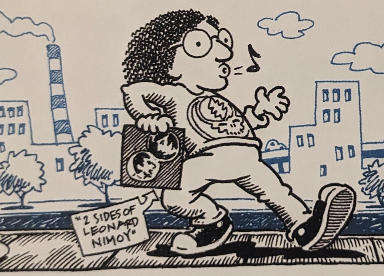
 @bourgwick@heads.social
grateful dead references in the summer issue of @…'s LCD programming guide
a.) the problems with record store day
b.) @… origin story!
c.) pfft, not even in the top 10 (& it's bitchin')
d.) full-page "mou…



 @AthanSpod@social.linux.pizza
I decided to try out Ecosia for search today, having been a long-time DuckDuckGo user.
Some observations:
1. For some bizarre reason there's no way to access your user profile on the main page, only on search results. No, not even if you create an account and login.
2. So once I'd found that... the searches are backed by Google and/or Bing. Thus I selected Bing to match my DDG experience.
3. They default to some form of AI enhancement, but at least you can t…
 @arXiv_qbioNC_bot@mastoxiv.page
[2026-07-21 Tue (UTC), 3 new articles found for q-bio.NC Neurons and Cognition]
toXiv_bot_toot
@arXiv_mathCT_bot@mastoxiv.page[2026-09-16 Wed (UTC), 3 new articles found for math.CT Category Theory]
toXiv_bot_toot
@arXiv_mathST_bot@mastoxiv.page[2026-08-17 Mon (UTC), 3 new articles found for math.ST Statistics Theory]
toXiv_bot_toot
@arXiv_astrophGA_bot@mastoxiv.pageCrosslisted article(s) found for astro-ph.GA. https://arxiv.org/list/astro-ph.GA/new
[3/3]:
- Physical Law Ecology: mapping multi-mechanism ecologies as the zeroth step of data-driven scienti...
Xiongheng Bian, Xiangyu Cui, Ma Feng, Xiaoyan Shen
https://arxiv.org/abs/2609.08536 https://mastoxiv.page/@arXiv_csSC_bot/117240025268605332
- Radial velocity follow-up of \textit{Gaia} astrometric substellar companions: six confirmed brown...
Marcus L. Marcussen, et al.
https://arxiv.org/abs/2609.08590 https://mastoxiv.page/@arXiv_astrophEP_bot/117240226771261973
- Compton-induced HE -- VHE $\gamma$-rays from pair cascade emissions in the SEDs of radio galaxies...
Mfuphi Ntshatsha, Markus B\"ottcher, Soebur Razzaque
https://arxiv.org/abs/2609.08787 https://mastoxiv.page/@arXiv_astrophHE_bot/117240359289783489
- Why Azimuthal Averaging Works in Halo Lensing: Symmetry and Power Counting for Nonlinear Shear an...
Keiichi Umetsu (ASIAA)
https://arxiv.org/abs/2609.08825 https://mastoxiv.page/@arXiv_astrophCO_bot/117240273367442521
- An analysis of four stellar rings
E. Paunzen, et al.
https://arxiv.org/abs/2609.08942 https://mastoxiv.page/@arXiv_astrophSR_bot/117240320949854065
- Open Clusters as Laboratories for Cool Star Evolution: Highlights from the Cool Stars 23 Splinter...
Deepak Chahal, et al.
https://arxiv.org/abs/2609.09026 https://mastoxiv.page/@arXiv_astrophSR_bot/117240331173721683
- Detection of episodic Ultra-fast Outflows in the Low-luminosity quasar Mrk 205 using XMM-Newton data
Victoria-Ceballos, Longinotti, Krongold, Lanzuisi, Gudi\~no, Lara-DI
https://arxiv.org/abs/2609.09028 https://mastoxiv.page/@arXiv_astrophHE_bot/117240376393874183
- TILING I: Field-level Bayesian reconstruction of cosmological initial conditions during the epoch...
Nikolaos Triantafyllou, Andrei Mesinger, Steven Murray, Samuel Gagnon-Hartman
https://arxiv.org/abs/2609.09102 https://mastoxiv.page/@arXiv_astrophCO_bot/117240294995454169
- Sizing the Universe with DESI Galaxy Sizes: Plain Fundamentals of Fundamental-Plane Lensing
Kun Xu, et al.
https://arxiv.org/abs/2609.09151 https://mastoxiv.page/@arXiv_astrophCO_bot/117240322128412863
toXiv_bot_toot
@arXiv_mathRA_bot@mastoxiv.pageReplaced article(s) found for math.RA. https://arxiv.org/list/math.RA/new
[1/1]:
- On axial algebras with $3$ eigenvalues
Vsevolod A. Afanasev
https://
@arXiv_csDS_bot@mastoxiv.pageA Strongly Subquadratic $(3 \varepsilon)$-Approximation for Weighted Edit Distance over Arbitrary Metrics
Ethan Mader, Borna Tavasoli, Jihan Wang
https://arxiv.org/abs/2609.14873
@arXiv_mathAT_bot@mastoxiv.pageReplaced article(s) found for math.AT. https://arxiv.org/list/math.AT/new
[1/1]:
- Relative cyclotomic structures and equivariant complex cobordism
Andrew J. Blumberg, Michael A. Mandell, Allen Yuan
https://arxiv.org/abs/2310.02348 https://mastoxiv.page/@arXiv_mathAT_bot/111180552474754562
- A multiplicative version of the tom Dieck splitting
Andrew J. Blumberg, Michael A. Mandell
https://arxiv.org/abs/2505.02721 https://mastoxiv.page/@arXiv_mathAT_bot/114459688814867416
- A rank-based distance for interval modules and Wasserstein stability of persistence landscapes
Peter Bubenik, Wanchen Zhao
https://arxiv.org/abs/2509.20921 https://mastoxiv.page/@arXiv_mathAT_bot/115269470873966833
- The rank-five Peterson hit problem, the Singer transfer, and canonical Milnor-operation layers
Dang Vo Phuc
https://arxiv.org/abs/2511.11745 https://mastoxiv.page/@arXiv_mathAT_bot/115569647485799776
- The Algebra of Categorical Spectra
Naruki Masuda
https://arxiv.org/abs/2605.03114 https://mastoxiv.page/@arXiv_mathAT_bot/116526500745660438
- Visualizing Local Maxima of the Ohio overdose epidemic with Vineyards
Nicholas Bermingham, David White, Nathan Willey
https://arxiv.org/abs/2607.05710 https://mastoxiv.page/@arXiv_mathAT_bot/116883198005698618
- Chern--Simons-type $3$-cocycles and $\mathbb{Q}/\mathbb{Z}$-torsion in the third homology of disc...
Takefumi Nosaka
https://arxiv.org/abs/2601.07230 https://mastoxiv.page/@arXiv_mathGT_bot/115886790889229457
- The Topology of $k$-Robust Clique Complexes in Grid-like Graphs
Marek Filakovsk\'y
https://arxiv.org/abs/2602.11365 https://mastoxiv.page/@arXiv_mathCO_bot/116062392661479542
- The Redshift Bound from Quillen-Lichtenbaum
Shay Ben-Moshe
https://arxiv.org/abs/2608.02180 https://mastoxiv.page/@arXiv_mathKT_bot/117036142171107624
toXiv_bot_toot
@arXiv_physicsinsdet_bot@mastoxiv.pageRadiotracer photoluminescence for element-specific identification of color centers
Brecht Biesmans, Afonso Lamelas, Kirill Danilov, Aleksandr Seliverstov, \^Angelo Costa, V\'itor Amaral, Andr\'e Vantomme, Jo\~ao Guilherme Correia, Ulrich Wahl, Lino M. C. Pereira, the ISOLDE Collaboration
https://arxiv.org/abs/2608.19219 https://arxiv.org/pdf/2608.19219 https://arxiv.org/html/2608.19219
arXiv:2608.19219v1 Announce Type: new
Abstract: We report on the implementation of a radiotracer photoluminescence spectroscopy setup at the ISOLDE radioactive ion beam facility at CERN, enabling element-specific identification of optically active defects in solids. The method combines radioactive ion implantation with optical spectroscopy, allowing the temporal evolution of photoluminescence signals to be correlated directly with nuclear decay. The setup is currently optimized for color centers in diamond and related wide-bandgap materials and enables room-temperature measurements. The system consists of an optical microscope coupled to a fiber-fed Czerny-Turner spectrometer with a liquid-nitrogen-cooled CCD detector, providing the stability required for long-duration measurements. As a proof-of-principle, radioactive $^{75}$Ga was implanted into diamond as a precursor to produce $^{75}$Ge impurities. The photoluminescence band extending from 600 nm, corresponding to the well-known GeV$^{-}$ center, exhibits an exponential decay with a half-life of $82.3^{ 2.5}_{-2.3} \mathrm{min}$, in agreement with the known $\beta^{-}$ decay half-life of $^{75}$Ge of $82.78(4) \mathrm{min}$. This establishes a direct and unambiguous correlation between the observed spectral feature and its germanium origin. These results demonstrate the capability of the setup to perform element-specific optical spectroscopy and extend radiotracer methods to color centers in wide-bandgap materials, taking advantage of the uniquely broad range of radioactive isotopes available at ISOLDE.
toXiv_bot_toot
@arXiv_physicsmedph_bot@mastoxiv.pagePRIME-SVR: Physics-infoRmed Implicit Multi-Echo Slice-to-Volume Reconstruction for Fetal T2 mapping
Busra Bulut, Maik Dannecker, Thomas Sanchez, Sara Neves Silva, Steven Jia, Jean-Baptiste Ledoux, Leo Pomar, Joanna Sichitiu, Yvan Gomez, Meriam Koob, Vincent Dunet, Maria Deprez, Guillaume Auzias, Francois Rousseau, Jana Hutter, Daniel Rueckert, Meritxell Bach Cuadra
https://arxiv.org/abs/2607.20136 https://arxiv.org/pdf/2607.20136 https://arxiv.org/html/2607.20136
arXiv:2607.20136v1 Announce Type: new
Abstract: Slice-to-volume reconstruction (SVR) is the standard method for obtaining high-resolution (HR) 3D fetal brain volumes from motion-corrupted 2D MRI slice stacks acquired in multiple orientations. Existing SVR methods are optimized and validated only for clinical-range echo times (TEs), limiting their use at non-clinical TEs and making them incompatible with quantitative T2 mapping, a protocol- and center-independent biomarker of fetal brain maturation requiring HR reconstructions across multiple TEs. We present PRIME-SVR, the first implicit neural representation (INR) framework for joint HR reconstruction from multi-echo MRI. A single fully connected network models a continuous function from spatial coordinates to signal intensities across TEs, while a second network estimates slice-specific acquisition degradations. Cross-TE coherence is enforced via a Bloch equation-derived regularization penalizing deviations from expected T2 decay, with adaptive weighting that strengthens coupling for degraded stacks. The method is fully self-supervised. We validate PRIME-SVR on 39 in vivo fetal acquisitions (13 subjects x 3 TEs) from two centers, two vendors, and two field strengths (1.5 T and 0.55 T). Compared to state-of-the-art SVR, PRIME-SVR improves reconstruction sharpness by 47%, anatomical accuracy by 30%, and cross-TE structural consistency by 14%. It enables reconstruction at late TEs previously inaccessible to SVR, yielding the first 0.8 mm isotropic T2 maps at 0.55 T and the first T2 maps derived from INR-based SVR. PRIME-SVR also accelerates quantitative imaging by reducing the data needed for multi-TE reconstruction, cutting acquisition from 15 to 10 minutes while keeping T2 accuracy within 1.7% in white and deep gray matter, or to 5 minutes with a mean T2 error of 2.3% for high-quality acquisitions.
toXiv_bot_toot
@arXiv_astrophGA_bot@mastoxiv.pageCrosslisted article(s) found for astro-ph.GA. https://arxiv.org/list/astro-ph.GA/new
[2/3]:
- Shaving the Outskirts of Planetary Systems Probed by Roman via Stellar Flybys
Donald Liveoak, Tim Hallatt, Sarah Millholland
https://arxiv.org/abs/2609.06112 https://mastoxiv.page/@arXiv_astrophEP_bot/117240099000374426
- PERRY: A Human-in-the-Loop GUI for Precision Alignment of MUSE Data
Bazkiaei, Miszalski, van de Sande, Lorente, O'Toole, Sheng
https://arxiv.org/abs/2609.06400 https://mastoxiv.page/@arXiv_astrophIM_bot/117240072263459997
- DustRover: A Python Package for Modelling Dust Extinction Curves (Phase I)
Amir E. Bazkiaei, Tayyaba Zafar, Nuria P. F. Lorente, Anilkumar Mailvaganam, Arihant Raidani
https://arxiv.org/abs/2609.06401 https://mastoxiv.page/@arXiv_astrophIM_bot/117240079505346895
- Chandra X-ray imaging and IC/CMB model for the inner jet of PKS 0637-752
Jaya Maithil, Daniel A. Schwartz, Aneta Siemiginowska, Diana M. Worrall, Preeti Kharb
https://arxiv.org/abs/2609.06587 https://mastoxiv.page/@arXiv_astrophHE_bot/117240161495404904
- Calibration of CNEOS Fireball Velocities and the Robustness of Nominally Hyperbolic Events
Volkan Duran, Abraham Loeb
https://arxiv.org/abs/2609.06745 https://mastoxiv.page/@arXiv_astrophEP_bot/117240133608812317
- Elusive helium stars in the gap between subdwarfs and Wolf-Rayet stars III. Formation of the Gala...
L. R. Yungelson, A. G. Kuranov, A. V. Mishakina
https://arxiv.org/abs/2609.06764 https://mastoxiv.page/@arXiv_astrophSR_bot/117240134556915599
- Little Red Dot Cosmology: A Matter-Era Baryon Acoustic Oscillations Probe of $\Lambda$CDM
Jessica A. Zebrowski, Rohan P. Naidu
https://arxiv.org/abs/2609.06926 https://mastoxiv.page/@arXiv_astrophCO_bot/117240064791330387
- An Eccentric Massive Protobinary Assembled via a Core-merger Parabolic Encounter
Yao Wang, et al.
https://arxiv.org/abs/2609.07390 https://mastoxiv.page/@arXiv_astrophSR_bot/117240144389587963
- Approximating the statistics of a gravitational wave background
Mikel Falxa
https://arxiv.org/abs/2609.07686 https://mastoxiv.page/@arXiv_astrophIM_bot/117240144585484923
- Precise physical and kinematical parameters of massive eclipsing binaries in the Small Magellanic...
M. Kaya, \"O. \c{C}ak{\i}rl{\i}, B. Hoyman, O. \"Ozdarcan, T. Yontan, R. Canbay
https://arxiv.org/abs/2609.07870
toXiv_bot_toot
@@arXiv_physicsatomph_bot@mastoxiv.page@mastoxiv.pageReplaced article(s) found for physics.atom-ph. https://arxiv.org/list/physics.atom-ph/new
[1/1]:
- Precision Measurement of the Saturation Intensity in Rubidium at 420 nm
Shivam Sinha, Sumit Achar, Sankar Satheesh, Arijit Sharma
@arXiv_physicsfludyn_bot@mastoxiv.pageTheoretical development of an operational wave-induced ice erosion model through laboratory experiments
Wenjun Lu, Behnam Ghadimi, Dominique Mouaze, Marianne Font, R\'emi Lambert, Harvey Goodwin, Widar Weizhi Wang, Raed Lubbad, Sveinung L{\o}set
https://arxiv.org/abs/2607.20118 https://arxiv.org/pdf/2607.20118 https://arxiv.org/html/2607.20118
arXiv:2607.20118v1 Announce Type: new
Abstract: Wave-induced melting of vertical ice fronts is represented in several operational iceberg and coastal-erosion models by the rough-wall parameterization of White (1980), whose closure chain is incompletely documented and whose commonly used compact expression is stated at the waterline. We reconstruct the formulation, specify the rough-turbulent wave-friction closure using Jonsson's implicit relation and its Lambert-W solution, and extend the model to a depth-resolved melt-rate profile under linear wave kinematics. Because the horizontal and vertical orbital-velocity components are linked, we use the horizontal component as a convenient representative scale and introduce a dimensionless coefficient alpha for the remaining closure uncertainty. The reconstruction gives a waterline coefficient of 3.0 x 10^-4 with White's resultant-velocity definition and 2.09 x 10^-4 for the reference choice alpha = 1; neither reproduces White's published 1.46 x 10^-4 directly. Two monochromatic wave-flume experiments with freshwater ice are then used to calibrate alpha from profiles below the wave trough. The full Lambert-W friction coefficient is used in this calibration. Best-fit values are 0.684 and 0.612 for periods of 1.54 and 0.87 s, respectively, a relative difference of approximately 11%. Their corresponding effective waterline coefficients, 1.43 x 10^-4 and 1.28 x 10^-4, are close to White's published value but do not constitute an independent validation. The fitted profiles reproduce the observed depth dependence below the trough, while deviations near the surface expose unresolved effects of intermittent submergence, local wave impact, and uncertain thermal forcing.
toXiv_bot_toot
@arXiv_mathDG_bot@mastoxiv.pageReplaced article(s) found for math.DG. https://arxiv.org/list/math.DG/new
[3/3]:
- A numerically flat rank-two bundle without a holomorphic connection on a $\partial\bar\partial$-t...
Tianzhi Hu, Runze Zhang
https://arxiv.org/abs/2609.08757 https://mastoxiv.page/@arXiv_mathCV_bot/117240229130532493
toXiv_bot_toot
@arXiv_hepth_bot@mastoxiv.pageReplaced article(s) found for hep-th. https://arxiv.org/list/hep-th/new
[3/3]:
- Distinct finite-temperature phase diagrams of non-invertible Kennedy--Tasaki duals
Weiguang Cao, Haruki Watanabe
https://arxiv.org/abs/2607.24231 https://mastoxiv.page/@arXiv_condmatstrel_bot/116996702950349969
- Renormalizations in holomorphic field theories on K\"ahler manifolds
Minghao Wang, Junrong Yan
https://arxiv.org/abs/2608.00546 https://mastoxiv.page/@arXiv_mathph_bot/117036123885412792
toXiv_bot_toot
@arXiv_astrophGA_bot@mastoxiv.pageCrosslisted article(s) found for astro-ph.GA. https://arxiv.org/list/astro-ph.GA/new
[1/3]:
- Exploring the dark sector through galaxy dynamics in clusters
Andrea Biviano, Lorenzo Pizzuti, Antonaldo Diaferio
https://arxiv.org/abs/2609.05597 https://mastoxiv.page/@arXiv_astrophCO_bot/117239937742870522
- The youngest white dwarf companion to a millisecond pulsar: Insights from NGC 362D
Ettorre, Dalessandro, Cadelano, Ridolfi, Pallanca, Freire, Krishnan, Salaris, D'Antona, Nag
https://arxiv.org/abs/2609.05608 https://mastoxiv.page/@arXiv_astrophSR_bot/117239974906161858
- Unresolved Binary Systems in the Rubin Era I: An Autoencoder Framework for Binary Identification ...
Tobias G\'eron, Alexander Laroche, Joshua S. Speagle, Maria R. Drout
https://arxiv.org/abs/2609.05616 https://mastoxiv.page/@arXiv_astrophSR_bot/117239984147520768
- Deep H$\alpha$ Imaging Survey of IC 348 with the Hubble Space Telescope: I. Accretion Properties ...
Jiang, Bowler, Zhou, Kraus, Andrews, Hillenbrand, Ireland, Zhu
https://arxiv.org/abs/2609.05620 https://mastoxiv.page/@arXiv_astrophSR_bot/117239988274911894
- Rapid radio variability in the $z\sim7$ blazar VLASS J0410$-$0139: Indication of a hidden populat...
Belladitta, Ba\~{n}ados, Gloudemans, Shimwell, Momjian, Intema, Mazzucchelli, Fendt, Vaidya, Walter
https://arxiv.org/abs/2609.05628 https://mastoxiv.page/@arXiv_astrophHE_bot/117240037069175396
- Calibration of the Next Generation Palomar Spectrograph as a Stellar Speedometer
Pranav Nagarajan, Kareem El-Badry
https://arxiv.org/abs/2609.05629 https://mastoxiv.page/@arXiv_astrophSR_bot/117240052598392711
- Unresolved triple systems in the aged open cluster Trumpler 19: Discovery, confirmation, and impl...
D. Minniti, et al.
https://arxiv.org/abs/2609.05722 https://mastoxiv.page/@arXiv_astrophSR_bot/117240070263744954
- A Uniform Census of Variable Stars in M101 Based on HST/ACS Photometry
Jiyu Wang, Xiaodian Chen, Pinjian Chen, Jianxing Zhang, Shu Wang, Licai Deng
https://arxiv.org/abs/2609.05825 https://mastoxiv.page/@arXiv_astrophSR_bot/117240081078431891
- Self-supervised reconstruction of transients in data from space-borne gravitational-wave detectors
Yuxiang Xu, Minghui Du, Bo Liang, Zihao Xiao, Tianyu Zhao, Peng Xu
https://arxiv.org/abs/2609.05890 https://mastoxiv.page/@arXiv_grqc_bot/117240126723091551
toXiv_bot_toot
@arXiv_mathST_bot@mastoxiv.pageConverse bounds for multiple graph alignment and correlation detection based on last matching
Taha Ameen, Bruce Hajek
https://arxiv.org/abs/2608.14450 https://arxiv.org/pdf/2608.14450 https://arxiv.org/html/2608.14450
arXiv:2608.14450v1 Announce Type: new
Abstract: The paper focuses on information theoretic converse bounds for the alignment of $m$ correlated graphs and for the detection of correlation among $m$ graphs. A simple idea for $m\geq 3$ is that if the alignment of $m-1$ of the graphs is revealed as extra information (by a genie for example) then it is still necessary to produce the alignment between the one remaining graph and the others, i.e. the last matching must be accomplished. For both Gaussian and Erdos-Renyi models, the last-matching problem is equivalent to one with two observed graphs, providing a path to extend converse bounds for $m=2$ to larger $m$. While the method is rather obvious for alignment, we show that the method can also be used to derive converse bounds for weak detection of correlation.
toXiv_bot_toot
@arXiv_mathCV_bot@mastoxiv.pageOn the exponential convergence of Kobayashi geodesics in strongly convex domains
Kingshook Biswas, Sanjoy Chatterjee
https://arxiv.org/abs/2607.17259 https://arxiv.org/pdf/2607.17259 https://arxiv.org/html/2607.17259
arXiv:2607.17259v1 Announce Type: new
Abstract: In this paper, we have proved a qualitative version of the approaching geodesic property for certain convex domains. More precisely, we have proved that that if $\Omega \subset \mathbb{C}^{d}$ is a bounded strongly convex domain with $\mathcal{C}^3$ boundary and $\gamma_{1}, \gamma_{2}:[0, \infty) \to \Omega$ are two geodesics such that $\gamma_{1}(\infty)=\gamma_{2}(\infty)=\xi \in \partial \Omega$. Then there exists $A\big(\gamma_{1}(0), \gamma_{2}(0) \big)>0$ and $T \in \mathbb{R}$ such that
$$K_{\Omega}(\gamma_{1}(t), \gamma_{2}(t T))\leq Ae^{-\frac{t}{2}} ~~~\,\hspace{1em} \forall t \geq 0.$$ Furthermore, using this property we provided a characterization of strongly pseudoconvex domain via a biholomorphic invariant function namely generalized squeezing function. We have proved that: For every $\alpha>2$ there exists $\epsilon(d,\alpha)>0$ such that the following holds: if $\Omega \subset \mathbb{C}^d$ is a bounded convex domain with $\mathcal{C}^{2,\alpha}$-boundary and \[ T_{\Omega}^{D}(z)\geq 1-\epsilon \] outside a compact subset of $\Omega$, where $D \Subset \mathbb{C}^{d}$ is a balanced strongly convex domain with $\mathcal{C}^{3}$ boundary and $T_{\Omega}^{D}$ is the squeezing function of $\Omega$ with respect to the domain $D$ then $\Omega$ is strongly pseudoconvex.
toXiv_bot_toot
@jon@henshaw.social @@arXiv_physicsatomph_bot@mastoxiv.page@mastoxiv.pageCrosslisted article(s) found for physics.atom-ph. https://arxiv.org/list/physics.atom-ph/new
[1/1]:
- Design of a Lamb-Shift Polarimeter for $^3$He Ions and Atoms
N. Faatz, R. Engels, C. Kannis, S. J. P\"utz, J. Steinhage
@arXiv_hepth_bot@mastoxiv.pageReplaced article(s) found for hep-th. https://arxiv.org/list/hep-th/new
[3/3]:
- Equivalence of Stability Criteria for Multi-Fluid Stars
Tian-Shun Chen, Xiao-Ding Zhou, Hao Feng, Kilar Zhang
https://arxiv.org/abs/2512.01640 https://mastoxiv.page/@arXiv_astrophHE_bot/115649670029097728
- ACT-Planck data and phase transitions from a viable no-scale Standard Model completion
Filippo Cutrona, Francesco Rescigno, Alberto Salvio
https://arxiv.org/abs/2603.24664 https://mastoxiv.page/@arXiv_hepph_bot/116300067793362249
- Rotation of the polarization plane in axion fields: application to neutron star polar cap regions
Iver H. Brevik, Moshe M. Chaichian, Tiberiu Harko, Yuri N. Obukhov
https://arxiv.org/abs/2603.28826 https://mastoxiv.page/@arXiv_hepph_bot/116328472387188451
- The scalar--Maxwell--$\Lambda(x)$ system: Wormhole spacetimes without nonlinear electrodynamics i...
G. Alencar, T. M. Crispim
https://arxiv.org/abs/2603.30003 https://mastoxiv.page/@arXiv_grqc_bot/116328672276108547
- Exact general relativistic solutions for a cylindrically symmetric stiff fluid matter source
Tiberiu Harko, Francisco S. N. Lobo, Man Kwong Mak
https://arxiv.org/abs/2604.02449 https://mastoxiv.page/@arXiv_grqc_bot/116356753476950318
- Gauged Q-balls in flat potentials
Julian Heeck, Yu Zhi
https://arxiv.org/abs/2604.07454 https://mastoxiv.page/@arXiv_hepph_bot/116379500037672008
- Electromagnetic, gravitational wave, and static gravitational transmission through throat spaceti...
Jeff Riley
https://arxiv.org/abs/2604.14238 https://mastoxiv.page/@arXiv_grqc_bot/116418938291963451
- Rotating black holes with primary hair in five-dimensional generalized Proca theory
Mokhtar Hassaine, Ulises Hernandez-Vera
https://arxiv.org/abs/2605.17870 https://mastoxiv.page/@arXiv_grqc_bot/116600369149996731
- Exact Entanglement Dynamics Beyond Nearest-Neighbor Dual-Unitary Floquet Systems
Tanay Pathak
https://arxiv.org/abs/2606.11311 https://mastoxiv.page/@arXiv_quantph_bot/116730376162758952
- Weyl gauge symmetry at LIGO-Virgo-KAGRA
D. M. Ghilencea, V. -M. Mandric
https://arxiv.org/abs/2607.13146 https://mastoxiv.page/@arXiv_grqc_bot/116928555436432190
- Two-Horizon Sector Thermodynamics as a Diagnostic for the Bi-Hair Organization of NUT Charge
Di Wu, Robert B. Mann, Shuang-Qing Wu
https://arxiv.org/abs/2607.23644 https://mastoxiv.page/@arXiv_grqc_bot/116996732836199490
toXiv_bot_toot
@@arXiv_physicsatomph_bot@mastoxiv.page@mastoxiv.pageReplaced article(s) found for physics.atom-ph. https://arxiv.org/list/physics.atom-ph/new
[1/1]:
- Ytterbium lattice clock with systematic uncertainty of $1.3\times 10^{-18}$ and instability at th...
Zhu, Shi, Zhang, Xu, Wang, Xiong, Xiong, Fang, Chen, He, Lyu
@arXiv_astrophGA_bot@mastoxiv.pageCrosslisted article(s) found for astro-ph.GA. https://arxiv.org/list/astro-ph.GA/new
[1/1]:
- Characterizing Microlensing Planetary System OGLE-2014-BLG-0676L with High-Resolution Image Const...
Asahi Idei, et al.
https://arxiv.org/abs/2607.18408 https://mastoxiv.page/@arXiv_astrophEP_bot/116962495878066247
- Nancy Grace Roman Space Telescope Wide Field Instrument: Bright Point Source Saturation Response ...
Dana R. Louie, et al.
https://arxiv.org/abs/2607.18419 https://mastoxiv.page/@arXiv_astrophIM_bot/116962545620205016
- Capture of interstellar objects during stellar encounters
Sean N. Raymond, Nathan A. Kaib, Matthew J. Hopkins
https://arxiv.org/abs/2607.18551 https://mastoxiv.page/@arXiv_astrophEP_bot/116962518094055064
- Semi-Analytical Model for the Evolution of Stellar Binaries in the Empty Loss Cone of Massive Bla...
Samuel McGuire, Evgeni Grishin, Ilya Mandel, Yuri Levin
https://arxiv.org/abs/2607.18761 https://mastoxiv.page/@arXiv_astrophHE_bot/116962539949026327
- Accretion of multipolar massive complex scalar field packets by a Schwarzschild black hole
Flavio Rosales-Infante, Ivan Alvarez-Rios, Francisco S. Guzman
https://arxiv.org/abs/2607.18920 https://mastoxiv.page/@arXiv_grqc_bot/116962643142466509
- A Type Ia Supernova Candidate at $z\sim4.3$: A Transient Interloper in the Search for $z\sim14$ G...
Seiji Toshikage, et al.
https://arxiv.org/abs/2607.18972 https://mastoxiv.page/@arXiv_astrophHE_bot/116962563120173845
toXiv_bot_toot
@arXiv_mathAT_bot@mastoxiv.pageReplaced article(s) found for math.AT. https://arxiv.org/list/math.AT/new
[1/1]:
- Representation characterization of equivariant geometric bordism
Hao Li, Bo Chen, Zhi L\"u, Qifan Shen
https://arxiv.org/abs/2501.06565 https://mastoxiv.page/@arXiv_mathAT_bot/113825435888761016
- On the integral cohomology of real toric manifolds
Feifei Fan
https://arxiv.org/abs/2509.06059 https://mastoxiv.page/@arXiv_mathAT_bot/115173630437550865
- Categorified Koszul duality of algebras
Isamu Iwanari
https://arxiv.org/abs/2512.12263 https://mastoxiv.page/@arXiv_mathAG_bot/115728435911956655
- $A_3$-formality for pro-2 Demushkin groups
Ambrus P\'al, Gereon Quick
https://arxiv.org/abs/2607.01028 https://mastoxiv.page/@arXiv_mathGR_bot/116849287251959106
- The Geometry of Cochains on Sampled Vietoris-Rips Complexes
Darrick Lee, Kelly Maggs
https://arxiv.org/abs/2608.11137 https://mastoxiv.page/@arXiv_mathDG_bot/117081537320356423
toXiv_bot_toot
@arXiv_physicsfludyn_bot@mastoxiv.pageA High-Order Flux Reconstruction Actuator-Line Framework for Rotating-Blade Aerodynamics on Fixed Cartesian Grids
Abdullah Al Imran, Meilin Yu
https://arxiv.org/abs/2607.20347 https://arxiv.org/pdf/2607.20347 https://arxiv.org/html/2607.20347
arXiv:2607.20347v1 Announce Type: new
Abstract: This work couples a high-order flux reconstruction/correction procedure via reconstruction (FR/CPR) solver with a rotating actuator-line model (ALM) to simulate rotating-blade aerodynamics on fixed Cartesian grids. Blade loading is represented by volumetric body force source terms projected through an isotropic Gaussian kernel in a blade-attached frame, eliminating the need to resolve blade geometry. Vertical-axis wind turbines (VAWTs) serve as the demonstration configuration, with a modified Boeing-Vertol dynamic stall model incorporated to capture unsteady lift and drag. A mesh-resolution criterion for the Gaussian projection kernel on reasonably coarse meshes is derived. It shows that cost-effective coarse meshes can operate in a mesh-controlled regime with negligible induction feedback, motivating a Double Multiple Streamtube (DMST) correction to recover the physical inflow. Simulations are carried out over a range of tip-speed ratios at a chord-based Reynolds number of Re_c ~ 3.6 x 10^5. The framework is validated against experimental near-wake measurements and previously reported LES-ALM results, and the mean wake profile shows good agreement. The predicted power-coefficient curve matches high-fidelity three-dimensional LES-ALM data to within 6% around the optimal VAWT operation conditions. The framework also captures the regime-dependent influence of dynamic stall, azimuthal blade loading, lift hysteresis, and characteristic wake structures. These results demonstrate that the FR/CPR-ALM framework provides an accurate and computationally efficient geometry-free approach for VAWT analysis, making it well suited for parametric studies and large-scale wind energy applications.
toXiv_bot_toot
@arXiv_mathCV_bot@mastoxiv.pageCertain real surfaces in $\mathbb{C}^2$ with degenerated CR singularities
Sushil Gorai, Suman Karak, Golam Mostafa Mondal
https://arxiv.org/abs/2607.17016 https://arxiv.org/pdf/2607.17016 https://arxiv.org/html/2607.17016
arXiv:2607.17016v1 Announce Type: new
Abstract: In this paper, we study the local polynomial convexity of certain smooth real surfaces in \(\mathbb{C}^2\) with isolated CR singularity at the origin with higher-order of degeneracy. Under the assumption that the surface can be pulled back to a union of finitely many pairwise transverse totally real surfaces by a proper holomorphic map from $\mathbb{C}^2$ to $\mathbb{C}^2$, we obtain a normal form for such surfaces near the origin as
$\{(z,w)\in\mathbb{C}^2: w= \overline{z}^k o(|z|^{k})\}$
or
$M_t
:=
\left\{
(z,w)\in\mathbb{C}^2 :
w=(z t\overline{z})^k o(|z|^k)
\right\}$,
for some \(t>0\), where the parameter $t$ is a local biholomorphic invariant. We focus on the surfaces with order of degeneracy $k\geq 3$.
We prove that $M_t$ is locally polynomially convex at the origin if $t>cosec\left(\frac{\pi}{k}\right)$.
On the other hand, for $0<\frac{1}{k-2}$, we will also show that $M_t$ fails to be locally polynomially convex at the origin; and furthermore, a $(2k-3)$-parameter family of analytic discs attached to $M_t$ for $0<\min\left\{\sin\left(\frac{\pi}{k}\right),\frac{1}{k-2}\right\}$.
toXiv_bot_toot
@arXiv_hepth_bot@mastoxiv.pageHolographic entanglement entropy with conformal boundary conditions
Elena C\'aceres, Hare Krishna, Harita Palani Balaji, Vaishnavi Patil
https://arxiv.org/abs/2608.06277 https://arxiv.org/pdf/2608.06277 https://arxiv.org/html/2608.06277
arXiv:2608.06277v1 Announce Type: new
Abstract: We study holographic entanglement entropy in 3-dimensional AdS gravity with conformal boundary conditions which fix the conformal class of the boundary metric and its extrinsic curvature, $K$, while leaving the Weyl mode dynamical. Extending Lewkowycz-Maldacena-Dong's replica construction, we derive the corresponding holographic entanglement entropy formula. We show that the fluctuating Weyl mode does not contribute additional entropy. The entropy of the full boundary is therefore the Bekenstein-Hawking entropy, and the entropy of a boundary subregion continues to obey the Ryu-Takayanagi prescription, namely, the area of a minimal surface divided by $4G_N$.
We carry out explicit calculations for global AdS, rotating and non-rotating BTZ geometries. For an interval in AdS$_3$ we find that the $K$-dependent holographic entanglement entropy is governed by $c_m=\frac{3 \ell}{2 G_N}.$ For a thermal state at high conformal temperatures we find that, the entropy is governed by $c_{\rm eff}=\frac{3 \ell}{2 G_N} \frac{K \ell- \sqrt{K^2 \ell^2-4}}{2}$, the same effective central charge that governs the Cardy-like density of states, in agreement with previous results in the literature.
Finally, we also compute the entanglement entropy directly from the conjectured dual boundary theory - a holographic CFT coupled with time-like Liouville theory and deformed by a marginal $T \bar{T}$ like operator - and find $S_{EE} =\frac{c_{\rm eff}}{3} \ln\!\left(\frac{2 R \sin\phi_0}{\epsilon}\right) .$ This result for the state with no operator insertions (vacuum state ), provides an independent boundary realization of $c_{\mathrm{eff}}$ while clarifying that this state is not the state dual to global AdS.
toXiv_bot_toot
@@arXiv_physicsatomph_bot@mastoxiv.page@mastoxiv.page[2026-07-21 Tue (UTC), 3 new articles found for physics.atom-ph Atomic Physics]
toXiv_bot_toot
@arXiv_mathDG_bot@mastoxiv.pageSingularities of the Wigner Caustic and the Centre Symmetry Set of Frontal Curves in the Euclidean Plane
Micha\l{} Zwierzy\'nski
https://arxiv.org/abs/2609.14653 https://arxiv.org/pdf/2609.14653 https://arxiv.org/html/2609.14653
arXiv:2609.14653v1 Announce Type: new
Abstract: Motivated by the role of the Wigner caustic in semiclassical phase-space analysis, we extend it, together with the centre symmetry set, from regular planar curves to cooriented frontals. For an angularly regular parallel pair, the signed speed of the Wigner caustic is one half of the difference of the extended signed radii of curvature, while singular points of the finite centre symmetry set are the critical points of their projective ratio. These formulas yield criteria and explicit invariants for ordinary and higher cusps, an order-lowering relation between the two constructions, and a multiplicity-weighted extension of the classical cusp-count inequality for strictly convex ovals.
We also analyse parallel pairs containing a singular frontal which is not a front. A $5/2$-cusp produces either a $5/2$- or, at a signed-radius resonance, a $5/3$-cusp on the Wigner caustic. The opposite resonance sends the centre symmetry set to infinity. We obtain the corresponding transfer results for a $5/3$-cusp and give a projective completion which resolves vanishing denominators and simultaneous finite-order zeros of the two signed radii.
toXiv_bot_toot
@arXiv_mathST_bot@mastoxiv.pageSharp proper estimation of fixed-component Gaussian location mixtures in polynomial time
Hengzhi He, Guang Cheng
https://arxiv.org/abs/2608.12701 https://arxiv.org/pdf/2608.12701 https://arxiv.org/html/2608.12701
arXiv:2608.12701v1 Announce Type: new
Abstract: We consider a mixture of at most $k$ unit-covariance Gaussians in $\mathbb{R}^d$ whose means belong to a fixed-radius ball, with no separation or minimum-weight condition. Doss, Wu, Yang and Zhou (2023) proved that the minimax Hellinger risk is of order $\sqrt{d/n}\wedge 1$ and constructed a proper polynomial-time estimator with the slower general bound $(d/n)^{1/4}$; obtaining the sharp rate in polynomial time for fixed $k\geq 3$ was left open. We resolve this question. The key device is a moment-fiber range finder. A second-moment subspace controls the energy missed by projection. We then estimate finitely many one-free-index Hermite contractions. These vector-valued contractions recover every tensor component containing exactly one missed direction at the sharp $\sqrt{d/n}$ scale. Every remaining term contains at least two missed factors and is therefore controlled by the residual second-moment energy. The resulting subspace has dimension depending only on $k$. Exhaustive moment fitting in this constant-dimensional space produces a proper mixture and, together with the dimension-free moment characterization of Gaussian mixtures, achieves the optimal Hellinger rate in polynomial arithmetic time for every fixed $k$.
toXiv_bot_toot
@arXiv_mathAT_bot@mastoxiv.pageCrosslisted article(s) found for math.AT. https://arxiv.org/list/math.AT/new
[1/1]:
- Obstructions to lifting quaternionic torus actions
Panagiotis Batakidis, Ioannis Gkeneralis
https://arxiv.org/abs/2608.07597 https://mastoxiv.page/@arXiv_mathDG_bot/117075727354491329
- Cycle-Decorated Ribbon Bar Complexes: Cut Factorization and Equivariant Homology
Pyuyi Chufeng Huang
https://arxiv.org/abs/2608.07599 https://mastoxiv.page/@arXiv_mathCO_bot/117075739542962366
- Topology of medial regime random simplicial complexes
Jonathan Ariel Barmak, Michael Farber
https://arxiv.org/abs/2608.07969 https://mastoxiv.page/@arXiv_mathCO_bot/117075954244315591
- Type Annihilation for Classifying Maps of Rack Spaces
Takefumi Nosaka
https://arxiv.org/abs/2608.08076 https://mastoxiv.page/@arXiv_mathGT_bot/117075791842906653
- Understanding Non-Split 2-Group Symmetry: (3 1)D SymTFT, Anomaly and Bordism
Zhenbang Gu, Ran Luo, Yi-Nan Wang, Yi Zhang
https://arxiv.org/abs/2608.08766
- On the internal homotopy theory of motivic categories
Dipankar Maity
https://arxiv.org/abs/2608.09592 https://mastoxiv.page/@arXiv_mathAG_bot/117075951295722696
toXiv_bot_toot
@arXiv_hepth_bot@mastoxiv.pageReplaced article(s) found for hep-th. https://arxiv.org/list/hep-th/new
[2/3]:
- Multicritical points of gravitational solitons and a black hole in four dimensions
Moaathe Belhaj Ahmed, Mois\'es Bravo-Gaete, Robert B. Mann, Constanza Quijada
https://arxiv.org/abs/2605.24783 https://mastoxiv.page/@arXiv_hepth_bot/116639897763402557
- Brane flows
Georgios Papadopoulos, Kostas Skenderis
https://arxiv.org/abs/2605.31181 https://mastoxiv.page/@arXiv_hepth_bot/116673829986872182
- Weyl conformal geometry vs Riemannian geometry of Weyl gauge invariant (dressed) metric
D. M. Ghilencea, V. -M. Mandric
https://arxiv.org/abs/2606.08080 https://mastoxiv.page/@arXiv_hepth_bot/116719153397467346
- Free-Field Construction of Heterotic String Compactified on Calabi-Yau Orbifolds via Corresponden...
Alexander Belavin, Doron Gepner, Grigory Makarov
https://arxiv.org/abs/2606.27490 https://mastoxiv.page/@arXiv_hepth_bot/116832327091097035
- Exact Planar Black Hole in AdS-Einstein-Scalar Gravity with IR-Emergent Nearly Conformal Fluid
Sangheon Yun
https://arxiv.org/abs/2606.31901 https://mastoxiv.page/@arXiv_hepth_bot/116843780286505261
- AdS Black Holes Are Short-Lived inside the Spectral Form Factor
Jos\'e L. F. Barb\'on, Eduardo Velasco-Aja
https://arxiv.org/abs/2607.21704
- Generalized comodule tube algebras for boundary and domain wall defects of (2 1)D topological order
Zhian Jia, Sheng Tan
https://arxiv.org/abs/2608.05071 https://mastoxiv.page/@arXiv_hepth_bot/117047570919781079
- Surviving correlations across a horizon: reflected entropy for bosonic fields in non-inertial fra...
Sayid Mondal
https://arxiv.org/abs/2608.05473 https://mastoxiv.page/@arXiv_hepth_bot/117053235449252443
- Cluster algebras and tilings for the m=4 amplituhedron
Even-Zohar, Lakrec, Parisi, Tessler, Sherman-Bennett, Williams
https://arxiv.org/abs/2310.17727 https://mastoxiv.page/@arXiv_mathCO_bot/111322217178993182
- Weak Correlations as the Underlying Principle for Linearization of Gradient-Based Learning Systems
Ori Shem-Ur, Khen Cohen, Aviv Orly, Yaron Oz
https://arxiv.org/abs/2401.04013
- Gravity and the Hierarchy Problem
Thede de Boer, Jisuke Kubo, Manfred Lindner, Markus Reinig
https://arxiv.org/abs/2510.12882 https://mastoxiv.page/@arXiv_hepph_bot/115382945122925880
toXiv_bot_toot
@arXiv_mathST_bot@mastoxiv.pageDensity Estimation on Compact Manifolds under Intrinsic Spectral Block Variation
Olga Klopp, Fedor Noskov
https://arxiv.org/abs/2608.12637 https://arxiv.org/pdf/2608.12637 https://arxiv.org/html/2608.12637
arXiv:2608.12637v1 Announce Type: new
Abstract: We introduce an intrinsic spectral sparsity model for nonparametric density estimation on compact connected Riemannian manifolds. Instead of penalizing coefficients in an arbitrarily chosen Laplace--Beltrami eigenbasis, we group each complete eigenspace and measure the Hilbert norm of its spectral component. The resulting block-variation space is basis independent and isometry invariant. We establish its structural, atomic, and nonlinear approximation properties and clarify its relation to Sobolev, Besov, and coefficientwise spectral $\ell^1$ classes. We then construct a coordinate-free block-shrinkage estimator and prove a nonasymptotic signal-dependent $L^2$-oracle inequality that adapts to the unknown set of detectable eigenspaces. Under polynomial spectral growth, the risk theory separates the number of spectral blocks from their multiplicities and exhibits two regimes: one driven by a single high-dimensional eigenspace and the other by cumulative spectral complexity. Under matching spectral-growth and nondegeneracy assumptions, corresponding minimax lower bounds show that this multiplicity dependence is intrinsic, with sharp consequences for spheres and the rotation group $SO(3)$. Finally, we develop a positive, normalized, block-penalized exponential spectral sieve for log-densities and derive likelihood oracle inequalities together with expected Kullback--Leibler, Hellinger, and $L^2$ risk bounds. The resulting framework provides a geometry-respecting theory of sparse density estimation that remains invariant under changes of eigenbasis.
toXiv_bot_toot
@arXiv_mathDG_bot@mastoxiv.pageA strict second-eigenvalue estimate for the scalar stability operator on surfaces in $\mathbb{RP}^{3}$
M\'arcio Batista, Abra\~ao Mendes
https://arxiv.org/abs/2608.05458 https://arxiv.org/pdf/2608.05458 https://arxiv.org/html/2608.05458
arXiv:2608.05458v1 Announce Type: new
Abstract: Let $\varphi:\Sigma^2\looparrowright\mathbb{RP}^3$ be a closed immersed surface, with no orientability or, equivalently, two-sidedness assumptions. We establish the sharp quantitative estimate $$ \lambda_2(\Delta |\sigma|^2 2)\leq2-\frac{2}{\operatorname{Area}(\Sigma)}\int_\Sigma H^2d\Sigma \frac{4\pi\chi(\Sigma)}{\operatorname{Area}(\Sigma)}. $$ Our main contribution is the analysis of the borderline case, which rules out equality when $\chi(\Sigma)\leq0$. More precisely, $$ \lambda_2(\Delta |\sigma|^2 2)<2\quad\text{whenever}\quad\chi(\Sigma)\leq0. $$ The proof combines the canonical Veronese embedding of $\mathbb{RP}^3$ into $\mathbb{S}^8$, the conformal test function method, an Obata-type rigidity argument, and the classification of closed flat minimal surfaces in $\mathbb{RP}^3$.
toXiv_bot_toot
@@arXiv_physicsatomph_bot@mastoxiv.page@mastoxiv.page[2026-08-19 Wed (UTC), 3 new articles found for physics.atom-ph Atomic Physics]
toXiv_bot_toot
@arXiv_mathAT_bot@mastoxiv.pageCrosslisted article(s) found for math.AT. https://arxiv.org/list/math.AT/new
[1/1]:
- The $\mathcal{L}$-Calculus for Causal Variational Principles: An Exterior Differential Calculus o...
Felix Finster, Niky Kamran, Frank van der Top
https://arxiv.org/abs/2608.08811 https://mastoxiv.page/@arXiv_mathph_bot/117075889558816751
- 3-dimensional TQFTs from derived categories of quantum group representations
Cris Negron
https://arxiv.org/abs/2608.10285 https://mastoxiv.page/@arXiv_mathQA_bot/117081418367597421
- $\mathrm{Mot}^{\mathrm{loc}}$ is not compactly generated
Maxime Ramzi
https://arxiv.org/abs/2608.10681 https://mastoxiv.page/@arXiv_mathKT_bot/117081422135401595
- The Geometry of Cochains on Sampled Vietoris-Rips Complexes
Darrick Lee, Kelly Maggs
https://arxiv.org/abs/2608.11137 https://mastoxiv.page/@arXiv_mathDG_bot/117081537320356423
toXiv_bot_toot
@arXiv_mathDG_bot@mastoxiv.pageRicci flow with metric torsion on surfaces of positive Euler characteristic
Shubham Dwivedi
https://arxiv.org/abs/2609.15880 https://arxiv.org/pdf/2609.15880 https://arxiv.org/html/2609.15880
arXiv:2609.15880v1 Announce Type: new
Abstract: We study an adapted Ricci flow of connections with metric torsion on surfaces with positive Euler characteristic. We first prove that there do not exist any nontrivial solitons of the flow on the $2$-sphere thus confirming a conjecture of Branding--Kr\"oncke (J. Geom. Anal. 27.3 (2017), arXiv:1606.09121). We give an explicit family of torsion data for which the corresponding global solutions fail to converge on $\mathbb{S}^2$. Nevertheless, we provide several sufficient conditions for the convergence of the flow to a stationary point. We first prove that the normalized adapted Ricci flow always converges on $\mathbb{RP}^2$, which completely answers a question in the paper of Branding and Kr\"oncke. Using this, we deduce that the flow converges on $\mathbb{S}^2$ whenever the initial metric and the torsion one-form are antipodally symmetric. We also prove a {\L}ojasiewicz--Simon gradient inequality for the flow and use it to prove convergence to a stationary point provided the solution is close to an arbitrary stationary point.
toXiv_bot_toot
@@arXiv_physicsatomph_bot@mastoxiv.page@mastoxiv.page[2026-09-18 Fri (UTC), 3 new articles found for physics.atom-ph Atomic Physics]
toXiv_bot_toot
@arXiv_hepth_bot@mastoxiv.pageReplaced article(s) found for hep-th. https://arxiv.org/list/hep-th/new
[3/3]:
- Distinct finite-temperature phase diagrams of non-invertible Kennedy--Tasaki duals
Weiguang Cao, Haruki Watanabe
@arXiv_astrophGA_bot@mastoxiv.pageReplaced article(s) found for astro-ph.GA. https://arxiv.org/list/astro-ph.GA/new
[3/3]:
- CSST large-scale structure analysis pipeline: IV. Cosmic Voids Identified from Galaxy Group Sampl...
Yingxiao Song, et al.
https://arxiv.org/abs/2606.01907 https://mastoxiv.page/@arXiv_astrophCO_bot/116679581913297366
toXiv_bot_toot
@@arXiv_physicsatomph_bot@mastoxiv.page@mastoxiv.page[2026-07-17 Fri (UTC), 3 new articles found for physics.atom-ph Atomic Physics]
toXiv_bot_toot
@arXiv_astrophGA_bot@mastoxiv.pageActive galactic nucleus activity in brightest cluster galaxies at intermediate redshifts in Sunyaev-Zel'dovich--selected clusters
N. Hatamkhani, R. E. Skelton, J. Delhaize, M. Hilton, S. I. Loubser, N. Gupta, K. M. Mogotsi
https://arxiv.org/abs/2609.09049 https://arxiv.org/pdf/2609.09049 https://arxiv.org/html/2609.09049
arXiv:2609.09049v1 Announce Type: new
Abstract: Context. Brightest cluster galaxies (BCGs) host powerful active galactic nuclei (AGNs) and help regulate the heating--cooling balance of the intracluster medium. While radio-mode feedback is well established locally, its prevalence at intermediate redshifts remains poorly constrained. Aims. We investigate the incidence, accretion state, and host-galaxy properties of AGNs in BCGs in Sunyaev--Zel'dovich (SZ)-selected clusters at $0.3 < z < 0.8$, testing dependences on cosmic epoch, cluster mass, and dynamical state. Methods. We analyse 171 BCGs from the Atacama Cosmology Telescope SZ catalogue, combining Southern African Large Telescope spectroscopy, WISE mid-infrared photometry, and Rapid ASKAP Continuum Survey radio data with \textsc{X-CIGALE} modelling to derive stellar masses, star formation rates, and Eddington-scaled accretion rates. Results. Thirty-eight BCGs (22%) host radio-loud AGNs. Only two ($\sim$5%) are high-excitation radio galaxies; the remainder are low-excitation systems. Most AGNs have low Eddington ratios ($-3 \lesssim \log_{10}\lambda_{\mathrm{Edd}} \lesssim -1$), consistent with radiatively inefficient accretion. Accretion efficiency increases with redshift, while correlations with cluster mass and dynamical state are weak. Radio luminosity is independent of cluster mass. Compared to field radio AGNs, BCG radio-loud AGNs have similar radio powers but are hosted by galaxies $\sim0.8$ dex more massive. Conclusions. BCG radio-loud AGNs at $0.3 < z < 0.8$ are predominantly low-excitation, radiatively inefficient systems, indicating that maintenance-mode accretion was widespread in massive clusters ($M_{500c} \simeq 3.2 \times 10^{14},M_\odot$) by $z \sim 1$. Accretion appears regulated primarily by fuel availability and cosmic evolution rather than global cluster properties, while the cluster environment mainly sets the host-galaxy mass scale.
toXiv_bot_toot
@arXiv_mathDG_bot@mastoxiv.pageLarge mass limits of $\mathrm{G}_2$ and Calabi--Yau monopoles: calibrated concentration, Higgs zeros, and abelianization
Daniel Fadel, Goncalo Oliveira
https://arxiv.org/abs/2608.05128 https://arxiv.org/pdf/2608.05128 https://arxiv.org/html/2608.05128
arXiv:2608.05128v1 Announce Type: new
Abstract: We study large mass monopoles with structure group $\mathrm{SU}(2)$ or $\mathrm{SO}(3)$ on asymptotically conical $\mathrm{G}_2$-manifolds and Calabi--Yau $3$-folds, with fixed asymptotic class. After placing the AC asymptotic theory, the variational compactness theory of Parise--Pigati--Stern, and Li's singular abelian compactness theory in a common $\Theta$-monopole framework, we prove that the mass-renormalized Yang--Mills--Higgs and intermediate energy measures converge to $8\pi\|T\|$ for a compactly supported calibrated integral codimension-three cycle $T$. This identifies the two limiting currents and shows that the variational calibration inequalities are saturated. Using this common limit as the starting point for a finer analysis, if $\mathcal S$ is the calibrated support, $\mathcal Z$ the Kuratowski upper limit of the Higgs zero sets, and $\mathcal C$ the limiting nonabelian locus, defined as the Kuratowski upper limit of Li's curvature concentration loci, then $\mathcal S\subset\mathcal Z\subset\mathcal C=\mathcal S\cup\mathcal O$, where $\mathcal O$ is precisely the obstruction to effective codimension-three monotonicity. For the cohomogeneity-one large mass families on the Bryant--Salamon $\mathrm{G}_2$-manifolds and the Stenzel Calabi--Yau $3$-fold, we prove that $\mathcal O=\varnothing$. On $X\setminus\mathcal C$ the sequence abelianizes; corrected longitudinal curvatures converge smoothly, and the remaining compactness alternatives are governed by $L^2$-harmonic $2$-forms.
toXiv_bot_toot
@arXiv_hepth_bot@mastoxiv.pageReplaced article(s) found for hep-th. https://arxiv.org/list/hep-th/new
[2/3]:
- Toward an Observable Algebra for de Sitter Space: Gap Protection and Modular Dressings
Hassan ElSayed
@arXiv_astrophGA_bot@mastoxiv.pageWeak cool core, weak turbulence: XRISM, XMM-Newton and Chandra spectroscopy of the core of Abell 1413
Egemen Gormez, Murat Hudaverdi
https://arxiv.org/abs/2609.08778 https://arxiv.org/pdf/2609.08778 https://arxiv.org/html/2609.08778
arXiv:2609.08778v1 Announce Type: new
Abstract: We present the first XRISM spectroscopy of the core of Abell 1413 (z = 0.143), combining 104.7 ks of Resolve with archival XMM-Newton and Chandra spectra of a matched sky region. A1413 is hot, massive and relaxed but lacks a strong cool core (t_cool = 6.4 Gyr), a regime in which the low turbulence reported to date has been tested only once, in A3571. The Resolve spectrum requires a single velocity-broadened component with kT = 7.06 ( 0.33, -0.43) keV and a line-of-sight velocity dispersion sigma_v = 170 ( 25, -24) km/s (M_3D = 0.217). The bulk velocity relative to the brightest cluster galaxy, v_bulk = -24 /- 26 (z_ICM) /- 71 (z_BCG) km/s, is limited by the optical redshift rather than by Resolve, and disfavours coherent line-of-sight sloshing. The non-thermal pressure fraction, 2.5 /- 0.7 per cent, implies a hydrostatic mass bias b < 2.5 per cent, comparable to the strong cool-core cluster A2029 (2.6 /- 0.3 per cent) and, at matched sampling scale, if anything below it. With A3571 (1.3 /- 0.3 per cent), to which A1413 rescales within 0.2 sigma, two weak cool cores now show that this quiescence does not require central cooling. The resolved Fe-K He-alpha complex gives Fe = 0.38 /- 0.03 (stat) /- 0.04 (sys) solar; Ni/Fe, separated from Fe K-beta for the first time, is solar, as is the pattern as a whole. The Ne, Mg and Si lines that set Ne/Fe, Mg/Fe and Si/Fe lie below 2 keV, where our instruments disagree by 10-20 per cent; we therefore do not regard the SNIa fraction as constrained.
toXiv_bot_toot
@arXiv_hepth_bot@mastoxiv.pageReplaced article(s) found for hep-th. https://arxiv.org/list/hep-th/new
[1/3]:
- Supersymmetry and Localization on Three-Dimensional Orbifolds
Matteo Inglese, Dario Martelli, Antonio Pittelli
@@arXiv_physicsatomph_bot@mastoxiv.page@mastoxiv.page[2026-08-14 Fri (UTC), 3 new articles found for physics.atom-ph Atomic Physics]
toXiv_bot_toot
@arXiv_astrophGA_bot@mastoxiv.pageJ023721.13$-$010528.5: A Giant $S$-shaped Radio Galaxy Showing Four Episodes of Jet Activity
Pavan Vijay Khadekar, Dharam Vir Lal, Ramana Athreya
https://arxiv.org/abs/2609.08506 https://arxiv.org/pdf/2609.08506 https://arxiv.org/html/2609.08506
arXiv:2609.08506v1 Announce Type: new
Abstract: We present high-sensitivity, high-resolution radio observations of J023721.13$-$010528.5 using the upgraded Giant Metrewave Radio Telescope in band-3 (250--500\,MHz) and band-4 (550--950\,MHz), along with MeerKAT (1280\,MHz) and VLA (3\,GHz) survey images. The radio source, hosted by a bright cluster galaxy at redshift $z=0.372$, is a giant radio galaxy with a projected linear size between the outermost hotspots of 1.2~Mpc and is one of the largest $S$-shaped radio sources known. Its radio morphology revealed four pairs of components straddling the radio core, which were also local maxima in profiles of radio flux densities and equipartition energy density measured along the radio lobes. Spatially resolved multi-frequency spectral analysis showed progressively older emission from the inner to the outer components, spanning approximately 4--21 Myr. The high degree of symmetry of the inner three component-pairs with respect to the radio core makes it improbable that these are randomly located knots in the radio jet. Therefore, we present J023721.13$-$010528.5 as the prototype of a new class of radio galaxies with four episodes of jet activity --- a quadruple-double radio galaxy. We suggest that giant $S$-shaped radio sources may be among the most promising targets for identifying and studying multi-epoch radio jet activity, since their large linear sizes and jet-axis evolution can spatially separate hotspots from successive episodes, making them easier to detect and analyse.
toXiv_bot_toot
@@arXiv_physicsatomph_bot@mastoxiv.page@mastoxiv.page[2026-09-10 Thu (UTC), 3 new articles found for physics.atom-ph Atomic Physics]
toXiv_bot_toot
@arXiv_astrophGA_bot@mastoxiv.pageVz-GAL Dusty Star-Forming Galaxies: Revisiting the CO-H2 Conversion Factor Tension
Prachi Prajapati, Axel Weiss, Dominik Riechers, Tom J. L. C. Bakx, Leindert A. Boogaard, Diana Ismail, Pierre Cox, Andrew J. Baker, Roberto Neri, Matthew Lehnert, Chentao Yang, Emilio Romano-Diaz, Hiddo S. B. Algera, Stefano Berta, Edoardo Borsato, Kirsty M. Butler, Asantha Cooray, Bethany Jones, Amelie Saintonge, Paul van der Werf
https://arxiv.org/abs/2607.18440 https://arxiv.org/pdf/2607.18440 https://arxiv.org/html/2607.18440
arXiv:2607.18440v1 Announce Type: new
Abstract: The CO luminosity-to-H$_2$ mass conversion factor ($\alpha_{CO}$) remains a debated uncertainty in determining molecular gas masses of high-redshift dusty star-forming galaxies (DSFGs). Dynamical mass constraints have often favored $\alpha_{CO}=0.8$~$M_{\odot}~{(K~km~{s}^{-1}~{pc}^{2})}^{-1}$, whereas dust- and radiative-transfer-based methods imply higher values. We revisit this ``tension" using the largest homogeneous sample of 21 unlensed $z\sim1-4$ DSFGs, with securely measured \coonezero luminosities from the VLA \vzgal survey and resolved ($\sim{0.1}^{\prime\prime}$) ALMA 1~mm dust continuum imaging. For 12 galaxies with robust modeling constraints, we derive molecular gas masses using dust spectral energy distribution modeling and the TUNER LVG framework, adopting a solar-metallicity gas-to-dust mass ratio of 100. Although not fully independent due to shared assumptions on dust properties, these approaches yield mutually consistent gas masses corresponding to $\alpha_{CO}\sim1.5-11.5$, with a median near the Galactic $\alpha_{CO}=4.3$. Isotropic virial dynamical masses agree with these gas masses when realistic molecular gas sizes are adopted, while our proposed ``mixed" (rotating, pressure-supported, thick-disk) estimator systematically underestimates dynamical masses, producing low $\alpha_{CO}$ limits. Using GN20 ($z=4.055$) as a case study, we show that resolved gas geometry and kinematics reconcile the discrepancy with LVG-derived $\alpha_{CO}$. Our results suggest that current data do not require $\alpha_{CO}=0.8$, and intermediate to near-Galactic values remain dynamically viable given uncertainties in gas geometry, dust properties, and gas-to-dust ratios. Further progress in calibrating $\alpha_{CO}$ in the early universe will require resolved molecular gas observations, physically motivated ISM modeling, and stringent constraints on dust properties.
toXiv_bot_toot
@@arXiv_physicsatomph_bot@mastoxiv.page@mastoxiv.pageDual-Platform Precision Measurement of the $3^2D_{5/2}$ to $4^2S_{1/2}$ $g$-Factor Ratio in $^{40}\text{Ca}^ $
Brian J. McMahon, Vikram S. Sandhu, John M. Gray, Creston D. Herold, Kenton R. Brown, Brian C. Sawyer
https://arxiv.org/abs/2607.07929
@arXiv_astrophGA_bot@mastoxiv.pageThe Next Generation Virgo Cluster Survey (NGVS). II. A Catalog of Galaxies in the Virgo Cluster
Laura Ferrarese, Patrick Cote, Lauren A. MacArthur, Joel C. Roediger, John P. Blakeslee, Michele Cantiello, Jean-Charles Cuillandre, Puragra Guhathakurta, Stephen Gwyn, Max M. Kurzner, Eric W. Peng, Matthew Santos, Eleanore B. Todd, Elisa Toloba, Pierre-Alain Duc, Patrick R. Durrell, Nicholas Fantin, Yuting Feng, Ariane Lancon, Sungsoon Lim, Chengze Liu, Deborah Lokhorst, Alessia Longobardi, Simona Mei, J. Christopher Mihos, Sanjaya Paudel, Thomas H. Puzia, Anand Raichoor, Ruben Sanchez-Janssen, Kaixiang Wang, Hongxin Zhang, Alessandro Boselli, Michael L. Balogh, Samuel Boissier, Eric Emsellem, J. J. Kavelaars, Alan W. McConnachie, Cameron R. Morgan, Chelsea Spengler, James E. Taylor, Matthew A. Taylor, Toby Brown, Sebastien Fabbro, Raphael Gavazzi, Hendrik Hildebrandt, Rashi Jain, Andres Jordan, Rory Smith, Solveig Thompson, Haruka Yoshino, Wim van Driel, Ludovic van Waerbeke, Tyrone E. Woods, Jade Yeung
https://arxiv.org/abs/2607.18414 https://arxiv.org/pdf/2607.18414 https://arxiv.org/html/2607.18414
arXiv:2607.18414v1 Announce Type: new
Abstract: The Next Generation Virgo Cluster Survey (NGVS) is a deep, high resolution imaging campaign that used the 1 deg$^2$ MegaCam instrument on the Canada-France-Hawaii Telescope to carry out a comprehensive optical survey of the Virgo cluster, from its core to its virial radius. The NGVS covers a contiguous area of 104 deg$^2$ (8.63 Mpc$^2$ at the 16.5 Mpc distance of Virgo) in the $u^*$-,$g$-,$i$-, and $z$-band, with additional limited coverage in $r$. In this paper, we present the final catalog of Virgo galaxies across the entire NGVS area. The catalog includes 3680 galaxies considered to be $bona~fide$ members of the cluster, spanning a factor of 2.5 million in luminosity, from $g = 8.42$ mag to $g = 24.41$ mag ($M_g = -22.67$ mag to $M_g = -6.68$ mag). With 2100 previously uncataloged galaxies, the NGVS catalog augments the number of known Virgo members by a factor 2.3. The catalog is complete down to $g = 18.6$ mag ($M_g=-12.5$ mag, corresponding to a stellar mass $M_* \sim 1.6\times10^7~M_{\odot}$ for an old stellar population) and 50% complete at $g = 22.0$ mag ($M_g=-9.1$ mag, $M_* \sim 6.2\times10^5~M_{\odot}$), three magnitudes deeper than the venerable Virgo Cluster Catalog (VCC), which for over 40 years has served as the reference standard for Virgo. Photometric and structural parameters are derived for all NGVS galaxies and presented in a series of tables, alongside nuclear and morphological classification, as well as stellar masses and, when available, radial velocities.
toXiv_bot_toot
@@arXiv_physicsatomph_bot@mastoxiv.page@mastoxiv.pageAbsolute frequency measurement of the $^{176}$Lu$^ \,(^{3}\mathrm{D}_1)$ standard against the NRC-FCs2 fountain with $2.6\times10^{-16}$ uncertainty
K. J. Arnold, Bin Jian, Zhao Zhang, Qi Zhao, Qin Qichen, N. Jayjong, M. D. K. Lee, Scott Beattie, M. D. Barrett
https://arxiv.org/abs/2607.07044
@@arXiv_physicsatomph_bot@mastoxiv.page@mastoxiv.pageAnalytical model for polarization transfer during gas-phase collision events in spin-exchange optical pumping: Spin-$\frac{1}{2}$ $^{129}$Xe versus spin-$\frac{3}{2}$ $^{131}$Xe
Perttu Hilla, Rajgowrav Cheenikundil, Juha Vaara
https://arxiv.org/abs/2609.04743