@jake4480@c.im
2026-06-28 20:52:20
New blog post on the importance of owning physical (and digital) media, when possible: https://spacetimetech.wordpress.com/2026/06/28/the-importance-of-owning-physical-and-digital-media-when-possible
@jake4480@c.imNew blog post on the importance of owning physical (and digital) media, when possible: https://spacetimetech.wordpress.com/2026/06/28/the-importance-of-owning-physical-and-digital-media-when-possible
 @lpryszcz@genomic.social
"Spotify's effective payout averages roughly $0.003 to $0.005 per stream... By contrast, Bandcamp pays artists roughly 82% of each sale. A direct $10 album sale can exceed revenue from thousands of streams. Buying music or books directly from artists, whether physical or DRM-free digital, is one of the most direct ways to support them."
Artists are likely better off when you #buy o…
 @cowboys@darktundra.xyz
Entire Cowboys roster passes physical, ready for camp today https://insidethestar.com/entire-cowboys-roster-passes-physical-ready-for-camp-today
 @Techmeme@techhub.social
Rockstar says physical copies of GTA 6 will contain a digital download code, not a disc; physical copies release on November 12, ahead of the November 19 launch (Tom Phillips/IGN)
https://www.ign.com/articles/grand-theft-auto-6…
 @davidaugust@mastodon.online
 @cdarwin@c.im
History repeats itself more than it should.
If you think we cannot get out from under these ghouls, then all I can say is we did it several times before.
We should be getting good at it by now.
The closest Trump's gonna get to heaven is on an airplane.
Go figure, cause I can't
Keep protests peaceful.
Don't kill anyone.
They DO make a difference.
Here are some resistance related guides from around the world:
🇺🇸 Fundamentals …
 @memeorandum@universeodon.com
Trump Undergoes a Physical Exam at Walter Reed (Karoun Demirjian/New York Times)
https://www.nytimes.com/2026/05/26/us/politics/trump-physical-walter-reed.html
http://www.memeorandum.com/260526/p63#a260526p63
@Techmeme@techhub.socialEliyan, which aims to license tech and make physical "chiplets" to ease AI chip data transfer bottlenecks, raised a $145M Series C at a $1B valuation (Stephen Nellis/Reuters)
https://www.reuters.com/business/eliyan-ra
 @bogo@hapyyr.com
I am challenging the status quo today (Jun-26) and submitted a physical (yes, offline) product at ProductHunt. Feel free to support me there!
#privacy
https://www.producthunt.com/products/paint
 @netzschleuder@social.skewed.de
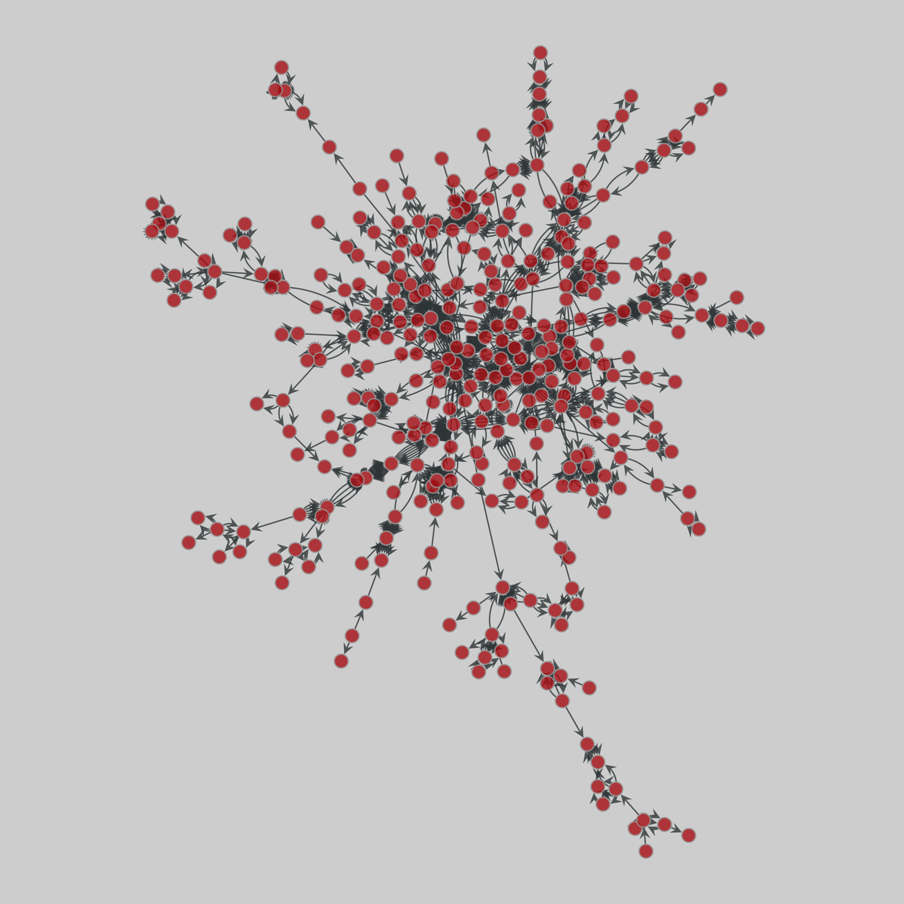
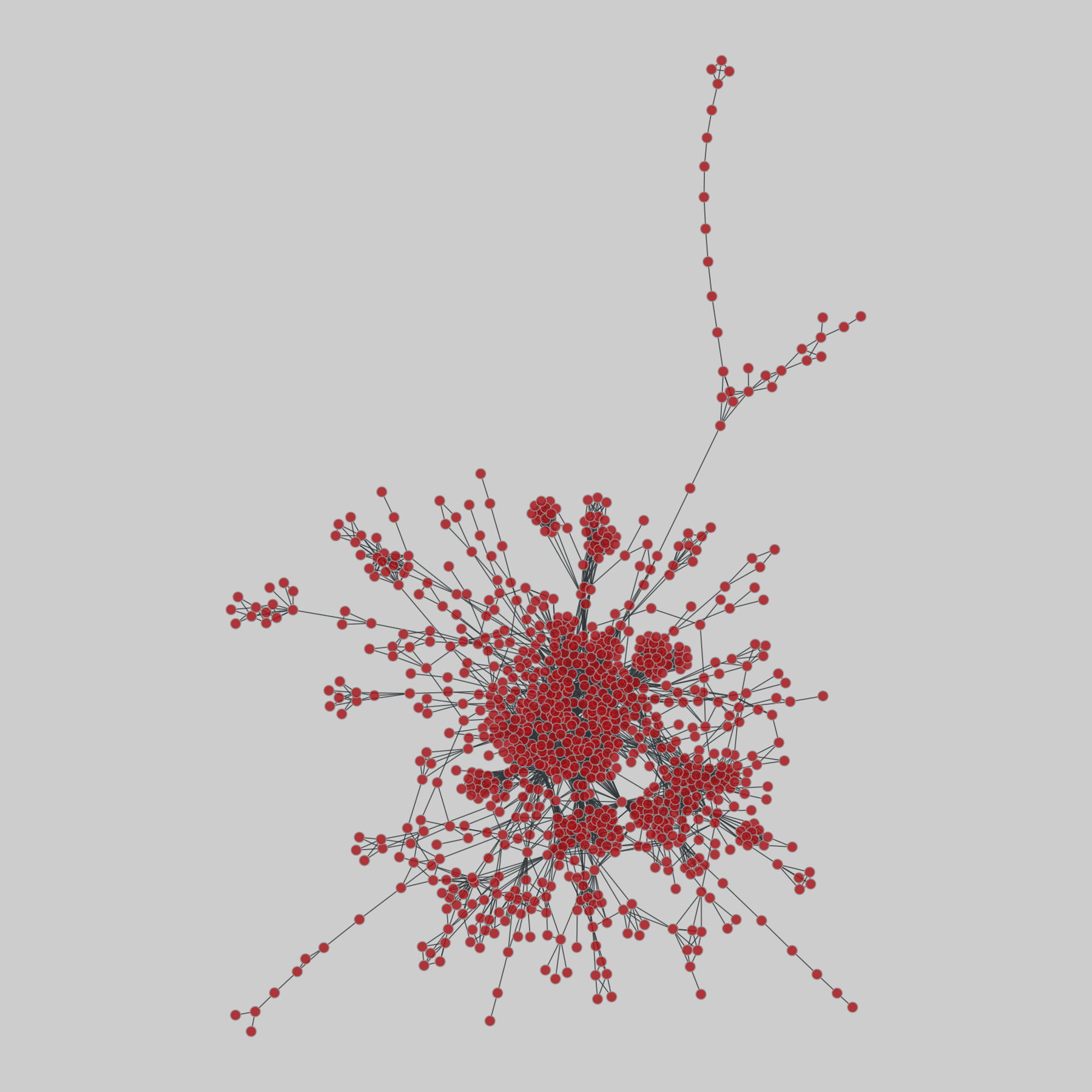
tree-of-life: Protein interactomes across the tree of life (2019)
Protein-protein iteraction networks ("interactome") for 1,840 species. An interactome captures all physical protein-protein interactions within one species, from direct biophysical protein-protein interactions to regulatory protein-DNA and metabolic interactions. .
This network has 610 nodes and 1988 edges.
Tags: Biological, Protein interactions
 @cdarwin@c.im
Since his first years as a top player, Jannik Sinner has long had one major weakness.
The hotter it is, the more vulnerable he becomes.
It nearly toppled him at January’s Australian Open, before Novak Djokovic did the job instead.
On Thursday, with temperatures climbing toward the 90s at Roland Garros, Sinner could not endure.
In the second round of the French Open, the world No. 1 fell victim to Juan Manuel Cerúndolo of Argentina, the world No. 56.
He grew dizzy…
 @raiders@darktundra.xyz
Eagles, Raiders Receive Compelling Maxx Crosby Message https://heavy.com/sports/nfl/las-vegas-raiders/eagles-compelling-message-maxx-crosby/
 @berlinbuzzwords@floss.social
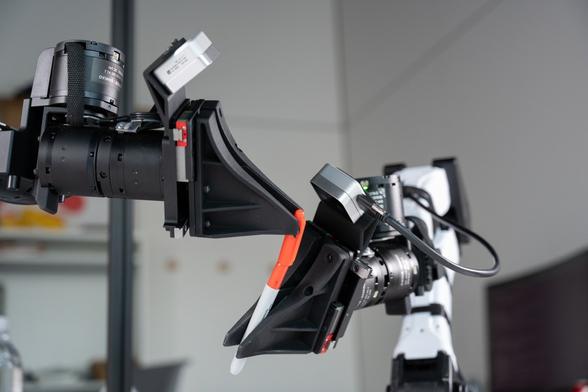
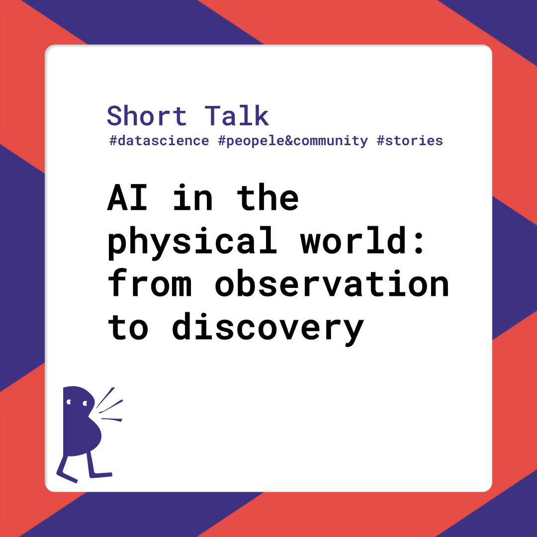
From collider experiments to large-scale astronomy systems, this session by Dmitriy Kostunin and Julian von Hoerschelmann-Schliwinski takes an honest look at where AI delivers measurable gains, and where it falls short.
Learn more about this session: https://2026.berlinbuzzwords.de/session/ai-in-the-physical-world-from-observation-to-discovery/
 @drgeraint@glasgow.social
Mathematical modelling of physical systems means deciding what features to include. I often advise my students that the very simplest models are the best place to start. E.g. start to model a car as a simple mass with forces applied to it, then add complexity later if needed, instead of getting bogged down by the details of every component at the start.
This FT article suggests a similar thing in an entertaining way.
£
@Techmeme@techhub.socialChipmaker Onsemi agrees to buy Synaptics in a nearly $7B all-stock deal expected to close in the middle of 2027; ON drops 9% and SYNA jumps 11% after hours (Samantha Subin/CNBC)
https://www.cnbc.com/2026/06/25/on-semi-synaptics-deal-physical-ai.html
…
 @bobmueller@mastodon.world
So what are your thoughts on these weighty book matters?
Yes
Long
"Real"/Physical
New
No, but it's not for lack of trying
#ThisOrThat
https://www.youtube.com/shorts/sHe_hqwpWaQ
@cdarwin@c.imThe Pentagon is moving to recruit hundreds of troops to appear as spectators at President Trump’s UFC cage-fighting event on the White House lawn,
and requiring those who attend to pay their own way and meet height and weight requirements.
https://www.
 @toxi@mastodon.thi.ng
New 10x8" silver gelatin print. This one took a lot of refining, back and forth and test strips to achieve the right balance between brightness, contrast (esp. in the brighter parts of the clouds) and smooth mid-tone transitions. So even though this is a contact print from a digital negative, this was the first time in ages (i.e. decades) that I used some physical dodging to soften the horizon and emphasize the haze in the distance a tiny bit more...


 @finlaydag33k@social.linux.pizza
>Remote for TV box (from ISP, not TV itself) misses physical buttons.
>Need to go to AI trashbot for "assistance"
>AI trashbot: "Have you tried turning the receiver off and on?"
>"The physical buttons are gone..."
>AI trashbot: "That can be caused by an empty battery, please replace them and let me know!"
 @NFL@darktundra.xyz
James Pearce Jr. returns to Falcons for minicamp physical amid intervention program https://www.nfl.com/news/james-pearce-jr-falcons-physical-mandatory-minicamp-intervention-program
@Techmeme@techhub.socialHuman Archive, which trains robots using first-person video from 1,000 camera-equipped caps worn by Indian home services workers, raised $8.2M from YC and more (Ivan Mehta/TechCrunch)
https://techcrunch.com/2026/05/26/human…
@netzschleuder@social.skewed.decopenhagen: Copenhagen Networks Study
A network of social interactions among university students within the Copenhagen Networks Study, over a period of four weeks, sampled every 5 minutes. Interactions include physical proximity (undirected), phone calls (directed, weighted), text messages (directed), and information about Facebook friendships (undirected). Nodes include some metadata, including gender.
This network has 536 nodes and 3600 edges.
Tags: Social, Offline, Unwei…
 @sean@scoat.es
A while back, I was mostly-lurking on a regional #Meshcore (like #Meshtastic , but different) discord, when someone on there tried to update a repeater and accidentally soft-bricked it. They needed to climb up to where it is to get a physical connection. But there's a raven nesting near where they've …
 @hikingdude@mastodon.social
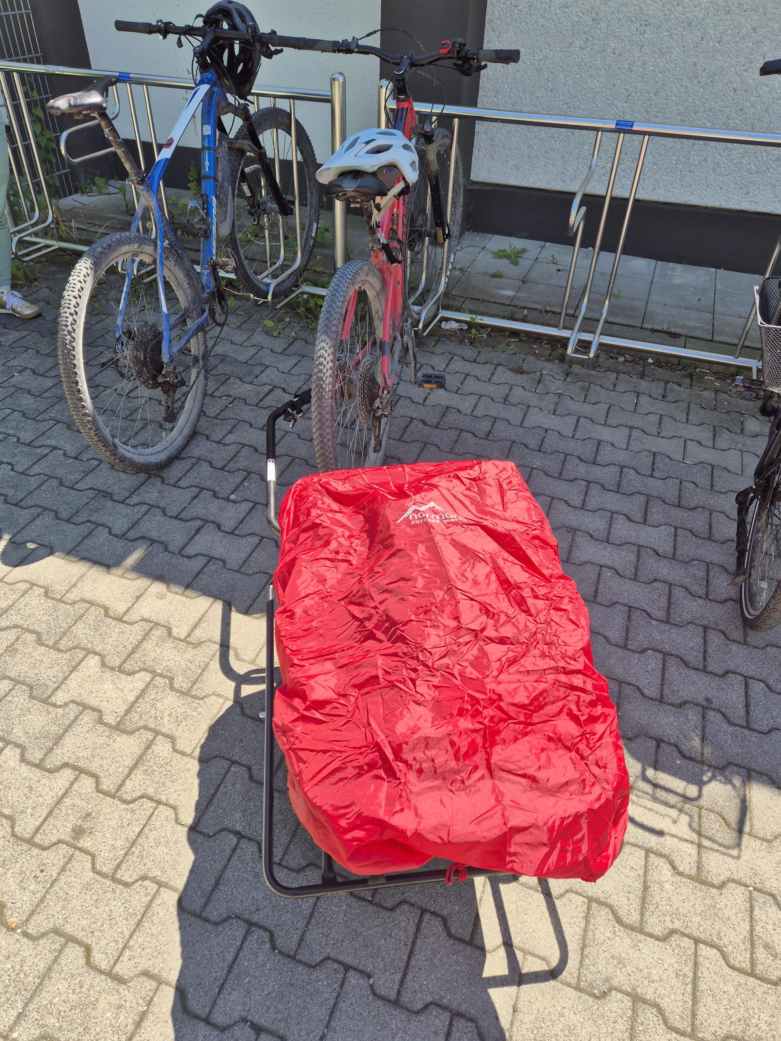
Yesterday we did our weekly errands again with our bicycle trolley. Works like a charm.
We select routes off major roads, have some chat along the way, see what's going on in town, physical exercise, fresh air, no fuel cost, no traffic jam.
And even car drivers are very friendly to us as well.
@Techmeme@techhub.socialOpenAI, Google, Meta, BlackRock, and others are recruiting electricians and carpenters by the thousands, with some of the highest pay the industry has ever seen (Lydia DePillis/New York Times)
https://www.ny…
 @KazuShuSora@social.tchncs.de
RE: https://mastodon.social/@Knoebel/116818598997578427
That's why you buy DRM-free / physical media 🙂↕️
 @cosmos4u@scicomm.xyz
"ESA is closely monitoring the wildfire situation in the vicinity of its deep-space ground station in #Cebreros, Spain," says https://www.esa.int/About_Us/Corporate_news/ESA_statement_on_the_wildfires_near_Cebreros: "Based on the information currently available, the station appears to have remained unharmed. [...] All ESA personnel are safe and accounted for. [...] At present, ESA does not have physical access to the station and is therefore unable to conduct an on-site inspection."
 @newsie@darktundra.xyz
Podcast: The Physical Politics of the Internet with Britt Paris https://www.404media.co/podcast-the-physical-politics-of-the-internet-with-britt-paris/
@davidaugust@mastodon.online @netzschleuder@social.skewed.decopenhagen: Copenhagen Networks Study
A network of social interactions among university students within the Copenhagen Networks Study, over a period of four weeks, sampled every 5 minutes. Interactions include physical proximity (undirected), phone calls (directed, weighted), text messages (directed), and information about Facebook friendships (undirected). Nodes include some metadata, including gender.
This network has 692 nodes and 2426279 edges.
Tags: Social, Offline, Un…
 @detondev@social.linux.pizza
I know next to nothing of substance abt The Amazing Digital Circus. All I know is that every time a trans woman makes a virtual reality story she makes a zillion bucks and her characters get turned into fortnite skins cuz of our innate sauce with the feeling of physical disconnection, and im lowk tempted to try continuing the family nacho recipe even as itd be purely cynical cuz I actually really really only care abt the process of becoming a real physical person im tired of thinking within …
 @Mediagazer@mstdn.social
The Guardian's tech team share their shoe-leather experiences covering AI data centers, as field investigation becomes increasingly important in tech coverage (Gautam Malkani/The Guardian)
https://www.theguardian.com/membership/202
 @david@boles.xyz
Three Books on a Table
When this novel began, there were three physical books on my table, and none of them had anything to do with the others....
https://bolesblogs.com/2026/07/24/three-books-on-a-table/
 @mszll@datasci.social
Why Your City Feels Built For Cars, Not People
https://www.forbes.com/sites/lauriewinkless/2026/07/24/why-your-city-feels-built-for-cars-not-people/
@memeorandum@universeodon.comTrump is seeing doctors for his annual physical. What the public finds out is up to him (STAT)
https://www.statnews.com/2026/05/26/trump-annual-physicial-medical-exam-public-scrutiny/
http://www.memeorandum.com/260526/p68#a260526p68
 @outer@mas.to
Wavefunction collapse is only one interpretation. Under some interpretations, graduate students also have souls.
#Comic

 @StephenRees@mas.to
From The Conversation
Where else can you peer into the labs and lecture halls of Canadian researchers and academics, who draw on their life's work to explain what being sedentary does to your body, why male chimps repeatedly throw rocks at trees and how the international legal order has collapsed?
We share their vast expert knowledge to educate Canadians so they can make informed decisions about politics, their health, the environment, the education of their children and the…
 @publicvoit@graz.social @cowboys@darktundra.xyz
Dallas Cowboys To Try More Physical Practice Approach For 2026 Training Camp https://www.si.com/nfl/cowboys/onsi/dallas-cowboys-to-try-more-physical-practice-approach-for-2026-training-camp
 @arXiv_physicsaoph_bot@mastoxiv.page
The physics of AI weather models
George Craig, Tobias Selz, Matthias Beylich, Kirsten I. Tempest
https://arxiv.org/abs/2605.23778 https://arxiv.org/pdf/2605.23778 https://arxiv.org/html/2605.23778
arXiv:2605.23778v1 Announce Type: new
Abstract: Could it be that AI weather models are solving physical equations, although they may not be the equations used by conventional NWP models? We compute correlations of forecast skill and Centered Kernel Alignment, providing evidence that different AI weather models represent the atmosphere in similar ways, despite differences in architecture and capacity. We argue that the architecture and training of the AI models constrains the form of the physical laws that they might simulate. In particular, we propose that the models implement a particle description of the atmosphere, where the latent variables at each mesh point correspond to the position of a particle in the high dimensional latent space. We hypothesize that the movement of the particles follows a gradient flow in the latent space towards a minimum of a learned free energy functional. Analysis of the GraphCast and Aurora models show that they make changes on large spatial scales in the early processor layers and move to smaller scale with increasing layer depth, consistent with the gradient flow hypothesis.
toXiv_bot_toot
 @nohillside@smnn.ch
 @mxp@mastodon.acm.org
“The project does not involve field research, laboratory experiments, or resource-intensive physical processes. The project-related activities are primarily computer-based and organizational in nature, and focus on the analysis and testing of data-driven infrastructures. Ecological sustainability aspects are therefore only marginally relevant.”
Well, the environmental impact of academic IT projects is indeed marginal compared to commercial operations, but…
⇢
@mxp@mastodon.acm.org“The project does not involve field research, laboratory experiments, or resource-intensive physical processes. The project-related activities are primarily computer-based and organizational in nature, and focus on the analysis and testing of data-driven infrastructures. Ecological sustainability aspects are therefore only marginally relevant.”
Well, the environmental impact of academic IT projects is indeed marginal compared to commercial operations, but…
⇢
 @metacurity@infosec.exchange
Physical attacks on cryptocurrency holders rose 75% in 2025, reaching 72 confirmed incidents and $41 million in known losses, according to data compiled by the blockchain security firm CertiK.
https://www.bloomberg.com/news/articles/20
@cdarwin@c.imI hear a longstandingly mentally ill man,
a narcissistic sociopath who is cognitively declining in his elder years,
becoming increasingly disinhibited.
We don't know exactly what his health is ...
He's obviously physically unwell.
— he's drooping on one side.
Did he have a left hemisphere CVA?
We don't know any of that, because they obviously would lie to us about any of it
 @arXiv_physicsmedph_bot@mastoxiv.page
Crosslisted article(s) found for physics.med-ph. https://arxiv.org/list/physics.med-ph/new
[1/1]:
- GPU-accelerated superiorization on constrained physical problems with SupPy
Tobias Becher, Yair Censor, Kay Barshad, Niklas Wahl
https://arxiv.org/abs/2606.27086 https://mastoxiv.page/@arXiv_physicscompph_bot/116815335801053799
toXiv_bot_toot
 @DamonHD@mastodon.social
 @grumpybozo@toad.social
I grew up with 2 sisters. I never heard either of them discuss guys in physical/visual terms.
My first girlfriend, later my first wife, said she was first attracted to me because I looked “like a man” whatever that meant. It only took her 5 years of intermittent dating & 10 of marriage to decide that maybe that was a poor basis.
My last girlfriend, later & still my second/last wife, hadn’t see a photo of me when she deemed me suitable from afar in `98
@cdarwin@c.imUS senator Mitch McConnell is still not cleared to return to work, his office confirmed on Monday,
six weeks after he was hospitalized after a fall at his home.
An update released alongside a new photograph of the Republican lawmaker – one of the highest-profile figures in Congress, as a former Senate majority leader – said he was undergoing “intense physical therapy” and is “working hard” to get back to his duties.
In the statement, he said that he wouldn’t be able to make…
 @thomasfuchs@hachyderm.io
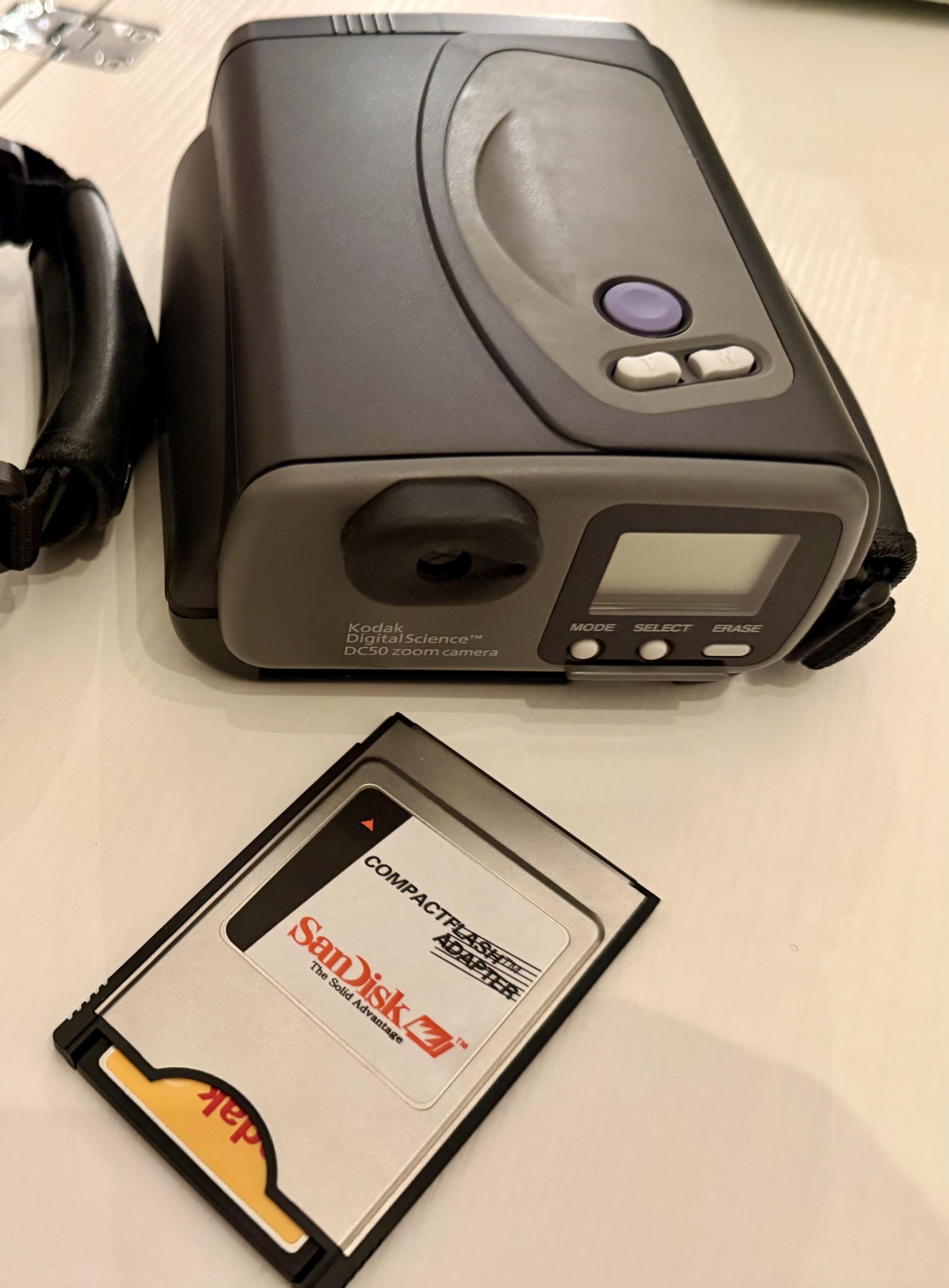
The quality of images from digital cameras is inversely proportional to the physical size of the memory card used.
This Kodak DC50 uses PCMCIA (type I or II) cards
 @danyork@mastodon.social @raiders@darktundra.xyz
Chargers mock Ravens failing Maxx Crosby physical in schedule release video https://raiderswire.usatoday.com/story/sports/nfl/raiders/2026/05/14/chargers-mock-ravens-failing-maxx-crosby-physi…
 @seav@en.osm.town
#TuneTuesday (May 19)
Lewis Capaldi uses his great voice to put out a bunch of hits and I like that he doesn’t let stardom get into his head and takes his mental and physical health seriously. Among his songs I’ve heard, “Forget Me” is easily my most favorite with his raspy vocals adding angst into the chorus.
 @realmurphy@social.linux.pizza
www.maptap.gg June 22
100🎯 91👑 98🎯 91👑 82🌟
Final score: 906
1. x: 4,686 pts, 97 km, 29 sec, 0 steps
2. x: 4,483 pts, 163 km, 25 sec, 0 steps
3. x: 498 pts, 3,441 km, 58 sec, 0 steps
4. x: 0 pts, 15,022 km, 53 sec, 0 steps
5. x: 0 pts, 13,783 km, 22 sec, 0 steps
Total: 9,667 pts, 32,506 km, 3 min, 8 sec, 0 steps
(out of physical time for the last two...)
TimeGuessr #1118 — 36,821/50,000
1️⃣ 🏆8666 - 📅4y - 🌍134.0 km
2️⃣ 🏆9908 - 📅0y…
@Techmeme@techhub.socialNvidia unveils Cosmos 3 Edge, a world model designed for robots and vision AI agents to perceive and navigate physical environments in real time (Jenny Lee/CNBC)
https://www.cnbc.com/2026/07/16/nvidia-reveals-new-ai-model-and-expands-…
@hikingdude@mastodon.socialDoing errands with our #suv (shopping utility vehicle). For the cover we bought the rain cover of an outdoor backpack. Works pretty well.
Meanwhile I also have an adapter mounted on my axel, too. so I can use it as well. 🎉
Best combination of shopping and physical activity. But I feel a little weird as hardly ever see anybody else with a trailer around. But I simply shouldn't care …
 @PwnieFan@infosec.exchange
For the second time in my life, I’m supporting a friend while they separate from an emotionally abusive partner. Both times, I didn’t even know there was abuse until the relationship ended. Here’s your PSA: Emotional abuse is just as damaging as physical abuse. And when a friend is suffering from it, they probably aren’t being noisy about it. They will quietly, slowly disappear from your life as their abuser convinces/cajoles/threatens them into abandoning who they are and what they love.
 @arXiv_physicsfludyn_bot@mastoxiv.page
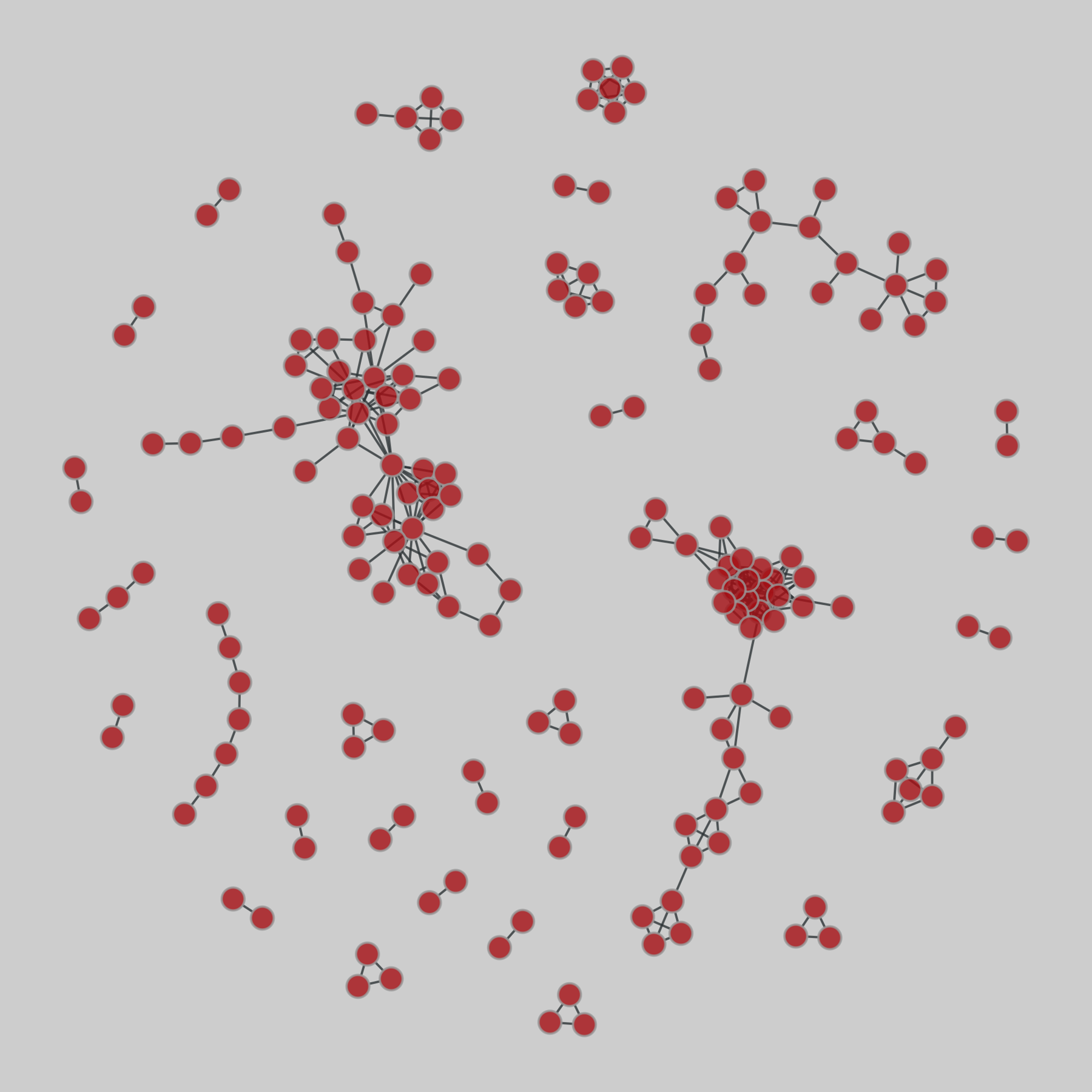
Parameter mapping and physical reconstruction of Akhmediev breathers at the interface of two fluid half-spaces
Olga Avramenko, Volodymyr Naradovyi
https://arxiv.org/abs/2607.20193 https://arxiv.org/pdf/2607.20193 https://arxiv.org/html/2607.20193
arXiv:2607.20193v1 Announce Type: new
Abstract: A methodology combining parameter mapping and physical reconstruction of Akhmediev breathers at the interface between two fluid half-spaces is developed on the basis of the Nayfeh model. Parameter maps are constructed to characterize the breather modulation period, the modulational instability growth rate, and the relative contribution of the bound second harmonic to the reconstructed interfacial profile. Their combined analysis provides a physically meaningful classification of breather regimes beyond the conventional focusing condition of the nonlinear Schr\"odinger equation. Reconstruction of the physical interfacial profile establishes a quantitative relation between nonlinear wave deformation and the contribution of the bound second harmonic, making it possible to identify the range of applicability of the weakly nonlinear approximation. Representative regimes from different modulational instability regions are analyzed to demonstrate the influence of resonance and focusing boundaries on breather characteristics and interfacial wave profiles. The proposed approach provides a direct link between the mathematical description of Akhmediev breathers and their physical interpretation and can be extended to other localized solutions of the nonlinear Schr\"odinger equation and to a broad class of stratified hydrodynamic systems.
toXiv_bot_toot
@Mediagazer@mstdn.socialWaPo's opinion section has been promoting data centers, only sometimes mentioning owner Jeff Bezos' vested interest in them or the paper's OpenAI partnership (Paul Farhi/Washingtonian)
https://washingtonian.…
@davidaugust@mastodon.online @cowboys@darktundra.xyzCan intriguing Cowboys UDFA back jump to front of line thanks to fit? https://cowboyswire.usatoday.com/story/sports/nfl/cowboys/2026/05/26/dominic-richardson-cowboys-scouting-report-udfa-2026/90256117007/…
@Techmeme@techhub.socialGermany's Black Forest Labs launches Flux 3 and Flux-mimic, its first models for robotics, as it expands from generative AI into physical AI (Yazhou Sun/Bloomberg)
https://www.bloomberg.com/news/articles/2026-0…
@NFL@darktundra.xyzNFL news roundup: Cowboys WR George Pickens reports for physical ahead of mandatory minicamp https://www.nfl.com/news/nfl-news-roundup-latest-league-updates-from-monday-june-15
@cosmos4u@scicomm.xyzUK astronaut #JohnMcFall could become the first person with a physical disability to live in orbit, thanks to an agreement signed between the UK government and US commercial space company #Vast: #Haven1 (not launched yet!) as early as next year.
@arXiv_physicsmedph_bot@mastoxiv.pageAdapting a 3D scanning water phantom for use in brachytherapy dosimetry
Rachael Wilks (Royal Brisbane and Women's Hospital, Herston Qld 4029, Australia, Herston Biofabrication Institute, Herston Qld 4029, Australia, University of Queensland, St. Lucia Qld 4072, Australia), Samuel C. Peet (Royal Brisbane and Women's Hospital, Herston Qld 4029, Australia, Herston Biofabrication Institute, Herston Qld 4029, Australia, Queensland University of Technology, Brisbane Qld 4001, Australia), Tanya Kairn (Royal Brisbane and Women's Hospital, Herston Qld 4029, Australia, Herston Biofabrication Institute, Herston Qld 4029, Australia, University of Queensland, St. Lucia Qld 4072, Australia, Queensland University of Technology, Brisbane Qld 4001, Australia), Scott B. Crowe (Royal Brisbane and Women's Hospital, Herston Qld 4029, Australia, Herston Biofabrication Institute, Herston Qld 4029, Australia, University of Queensland, St. Lucia Qld 4072, Australia, Queensland University of Technology, Brisbane Qld 4001, Australia)
https://arxiv.org/abs/2606.21320 https://arxiv.org/pdf/2606.21320 https://arxiv.org/html/2606.21320
arXiv:2606.21320v1 Announce Type: new
Abstract: In external beam radiotherapy, 3D scanning water phantoms are the gold standard for obtaining relative dosimetry data. These phantoms, consisting of a water tank and mechanical arm, along with accompanying software, are de-signed to acquire dose profiles along and orthogonal to the beam axis. In brachy-therapy, the acquisition of analogous dose profiles is more difficult, and is generally achieved with complex custom-built phantoms or chemical dosimeters such as film or gel. In this study, a low-cost 3D-printed jig was designed and fabricated within a clinical department, to allow precise brachytherapy dose measurements using a PTW BeamScan water phantom. Specifically, this jig al-lowed applicators to be suspended securely and reproducibly within the water phantom. Protocols were developed to relate the scanning system coordinates to the physical source position, and to obtain isodose planes both parallel and radial to the source axis. The developed solution has the potential to be used for physical verification of TG43 dose calculation parameters (e.g. anisotropy functions), the characterization of dose for a single dwell position in a complex applicator containing non-water equivalent materials, or the collection of point dose measure-ments for treatments incorporating multiple dwell positions or catheters.
toXiv_bot_toot
@arXiv_physicsaoph_bot@mastoxiv.pageLagged sea-surface-temperature precursors of the leading PM2.5 mode in China
Yuan Chen, Dan Zhao, Xu Li
https://arxiv.org/abs/2605.25436 https://arxiv.org/pdf/2605.25436 https://arxiv.org/html/2605.25436
arXiv:2605.25436v1 Announce Type: new
Abstract: Fine particulate matter(PM2.5) pollution in China is strongly modulated bymeteorological variability, yet its seasonal predictability from oceanic signals remains unclear. Here we identify the leading PM2.5 variability mode over China and show that it is preceded by coherent sea-surface-temperature anomaly clusters by more than one season. These oceanic precursors influence summer PM2.5 mainly by altering precipitation and lowlevel ventilation, and winter PM2.5 by modulating boundary-layer height and near-surface stagnation. Using the four largest precursor regions, a simple regression model achieves significant independent prediction skill for both summer and winter PM2.5 variability. Our results reveal a physical pathway linking sea-surface-temperature memory to regional aerosol pollution and provide a basis for seasonal air-quality risk assessment.
toXiv_bot_toot
@cdarwin@c.imPoetry gives people a language to express collective grief.
In Gaza, poetry documents what cameras cannot always reach and what numbers can never explain.
When destruction erases physical spaces, poetry becomes a witness to history.
https://w…
@Techmeme@techhub.socialSan Diego-based Aether AI, which is building "causal world models" to teach robots cause and effect instead of pattern-matching, raised a $20M seed led by MPCi (Cristian Dina/The Next Web)
https://thenextweb.com/news/aether-ai-causal-world-models-20m-…
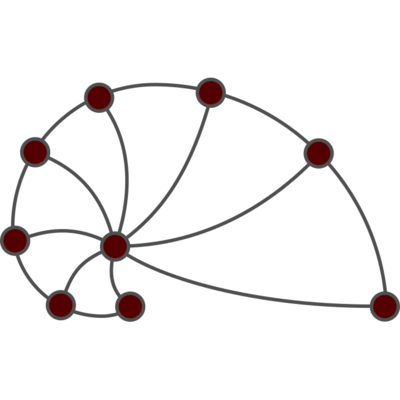
@netzschleuder@social.skewed.detree-of-life: Protein interactomes across the tree of life (2019)
Protein-protein iteraction networks ("interactome") for 1,840 species. An interactome captures all physical protein-protein interactions within one species, from direct biophysical protein-protein interactions to regulatory protein-DNA and metabolic interactions. .
This network has 186 nodes and 361 edges.
Tags: Biological, Protein interactions
 @jake4480@c.im
Mid year, trying to knock out a few blog posts. Finished the one on the Gemini protocol, since I wanted to do that one before I finish the small web one. Next up is a brief one on physical media, then the one on the small web after that. And maybe get a few more in after all those before the end of the year.
#blogs #blogging
@cdarwin@c.imPrometheus,
the physical AI startup co-founded by Jeff Bezos and Vik Bajaj, the former co-founder of Verily, Google’s life sciences unit,
announced it raised $12 billion at a $41 billion valuation.
The new funds came from Bezos,
as well as from JPMorgan Chase, Goldman Sachs, and BlackRock, among others.
This is the second fundraise round for Prometheus, which launched late last year with an initial raise of $6.2 billion, according to CNBC.
Prometheus is bu…
@Techmeme@techhub.socialElio, which is developing a new type of image sensor designed for AI rather than human vision, raised a $21M Series A led by Innovation Endeavors and Xora (Meir Orbach/CTech)
https://www.calcalistech.com/ctechnews/article/rkhattyhzl
@arXiv_physicsaoph_bot@mastoxiv.pageSeeing Inside the Storm: Improving Nowcasting by Integrating Meteorological Drivers
Minghui Qiu, Jun Chen, Lin Chen, Weifeng Chen, Shuxin Zhong, Zhidan Liu, Yu Zhang, Kaishun Wu
https://arxiv.org/abs/2605.24067 https://arxiv.org/pdf/2605.24067 https://arxiv.org/html/2605.24067
arXiv:2605.24067v1 Announce Type: new
Abstract: Most nowcasting systems, built on radar reflectivity, focus on current precipitation, ignoring the atmospheric precursors -- such as low-level convergence, turbulent eddies, and latent heating -- that offer a fleeting window to foresee storm birth. We introduce MeteoLogist, a physics-inspired radar intelligence framework that models the full life cycle of convection -- from its precursors to organized storm evolution. However, exploiting these precursors is non-trivial: they originate from multiple meteorological drivers -- thermodynamic, kinematic, and microphysical -- that evolve asynchronously (C1) and remain spatially fragmented (C2). To this end, MeteoLogist designs three tightly integrated components. The Physics-Tailored Encoders process radar echoes according to their intrinsic physical scales and semantics, forming thermodynamic, kinematic, and microphysical streams that capture distinct dynamical regimes. The Temporal-Phase Aligner addresses C1 by leveraging causal temporal attention to capture when and how different drivers interact and activate. The Cross-Field Spatial Aggregator addresses C2 through cross-regional fusion, aligning weak and scattered precursors across neighboring cells to expose upstream triggers and enforce spatial coherence. Evaluated on 3D-NEXRAD (2020--2022, US-wide), MeteoLogist boosts high-impact detection (CSI40) by 9.7% over strong baselines, and achieves a remarkable 37.67% gain during the storm-developing stage -- demonstrating true foresight in sensing storms before they appear. The code can be found in the supplementary material.
toXiv_bot_toot
@cowboys@darktundra.xyzCowboys' Brian Schottenheimer hints at more physical training camp ahead https://www.sportingnews.com/ca/nfl/dallas-cowboys/news/cowboys-brian-schottenheimer-padded-practices-oxnard/e00c3a30ef702b47e58df24b…
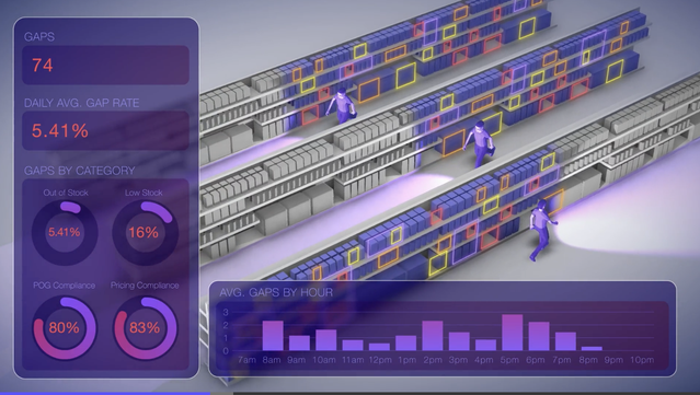
@Techmeme@techhub.socialSeattle-based Augmodo, whose AI-powered "Smartbadges" worn by employees track shelf inventory, raised $21M led by TQ Ventures at a $350M valuation (Kurt Schlosser/GeekWire)
https://www.geekwire.com/2026/augmodo-rais
@memeorandum@universeodon.comWhite House Releases Results of Trump's Latest Physical Exam (Chris Cameron/New York Times)
https://www.nytimes.com/2026/05/30/us/politics/trump-health-medical-physical-exam.html
http://www.memeorandum.com/260530/p43#a260530p43
@cdarwin@c.imLast year, we covered how Anthropic was buying physical books,
scanning them,
and destroying them to feed their Claude chatbot.
Judge William Alsup ruled this fair use.
In particular, he said mulching the books was perfectly fine because making them digital was
“exceedingly transformative”: [Order on Fair Use, 2025, PDF]
The print original was destroyed.
One replaced the other.
And, there is no evidence that the new, digital copy was shown, s…
@Techmeme@techhub.socialArm EVP Mohamed Awad says its chip architecture now accounts for 50% of the hyperscale cloud computing market, as AI demand transforms the data center industry (Cheng Ting-Fang/Nikkei Asia)
https://asia.nikkei.com/editor-s-picks
@arXiv_physicsmedph_bot@mastoxiv.pageOn the link between optoacoustic imaging and selective photothermolysis
Sergio Contador, Rodrigo Rojo, Alvaro Jimenez, Juan Aguirre
https://arxiv.org/abs/2606.25913 https://arxiv.org/pdf/2606.25913 https://arxiv.org/html/2606.25913
arXiv:2606.25913v1 Announce Type: new
Abstract: Selective photothermolysis (SP) is widely used in clinical and cosmetic dermatology to remove unwanted skin structures. Careful laser parameter selection results in safe and effective target removal. Nevertheless, parameter selection relies on a trial-and-error process based on visual inspection of the immediate skin response. This process is highly dependent on the practitioner experience and can be time-consuming.
SP and optoacoustic imaging (OI) share many physical principles. However, the possibility of using OI to improve laser parameter selection in SP has not been studied before. Here, we explore the relationship between OI and SP theoretically and through clinical in-human trials with a focus on tattoo removal. Our results demonstrate a strong correlation between OI signals acquired before and after treatment with the immediate clinical endpoint, suggesting that OI could be used as a tool for optimal parameter selection and reduced treatment duration in tattoo removal and other SP treatments.
toXiv_bot_toot
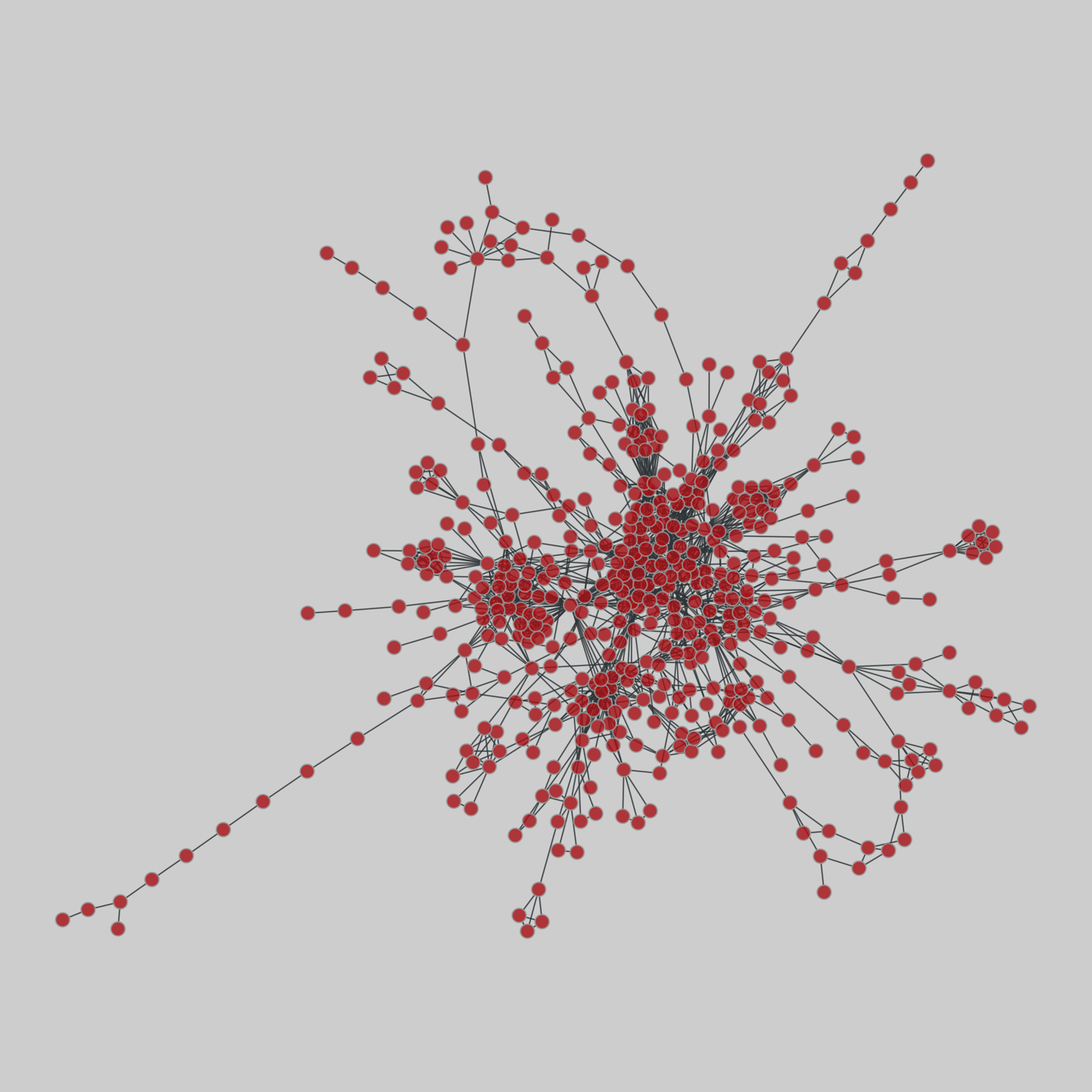
@davidaugust@mastodon.online @netzschleuder@social.skewed.detree-of-life: Protein interactomes across the tree of life (2019)
Protein-protein iteraction networks ("interactome") for 1,840 species. An interactome captures all physical protein-protein interactions within one species, from direct biophysical protein-protein interactions to regulatory protein-DNA and metabolic interactions. .
This network has 1369 nodes and 6629 edges.
Tags: Biological, Protein interactions
 @Techmeme@techhub.social
Alibaba's AI unit Tongyi Lab launches the Qwen Robot Suite, its first family of AI models for robots, in pilot testing with some enterprise clients (Wency Chen/South China Morning Post)
https://www.scmp.com/tech/big-tech/article
@Techmeme@techhub.socialGoogle plans to open its first physical Google Store outside of the US, in Tokyo's Omotesando district "this summer", marking Google's 11th physical store (Damien Wilde/9to5Google)
https://9to5google.com/2026/06/01/goog
@Techmeme@techhub.socialIntel taps Alex Katouzian, an ex-Qualcomm EVP, to lead Client Computing & Physical AI group, and names Pushkar Ranade as CTO, after serving on an interim basis (Dylan Martin/CRN)
https://www.crn.com/news/components-periph
@davidaugust@mastodon.online @cdarwin@c.imA U.S. appeals court has ruled in a case involving Exxon Mobil that the U.S. Occupational Safety and Health Administration
cannot require businesses to document work-related mental illnesses reported by employees.
A unanimous three-judge panel of the New Orleans-based 5th U.S. Circuit Court of Appeals said on Tuesday
that a requirement in federal law that employers periodically report work-related deaths, injuries and illnesses only applies to physical -- and not mental c…
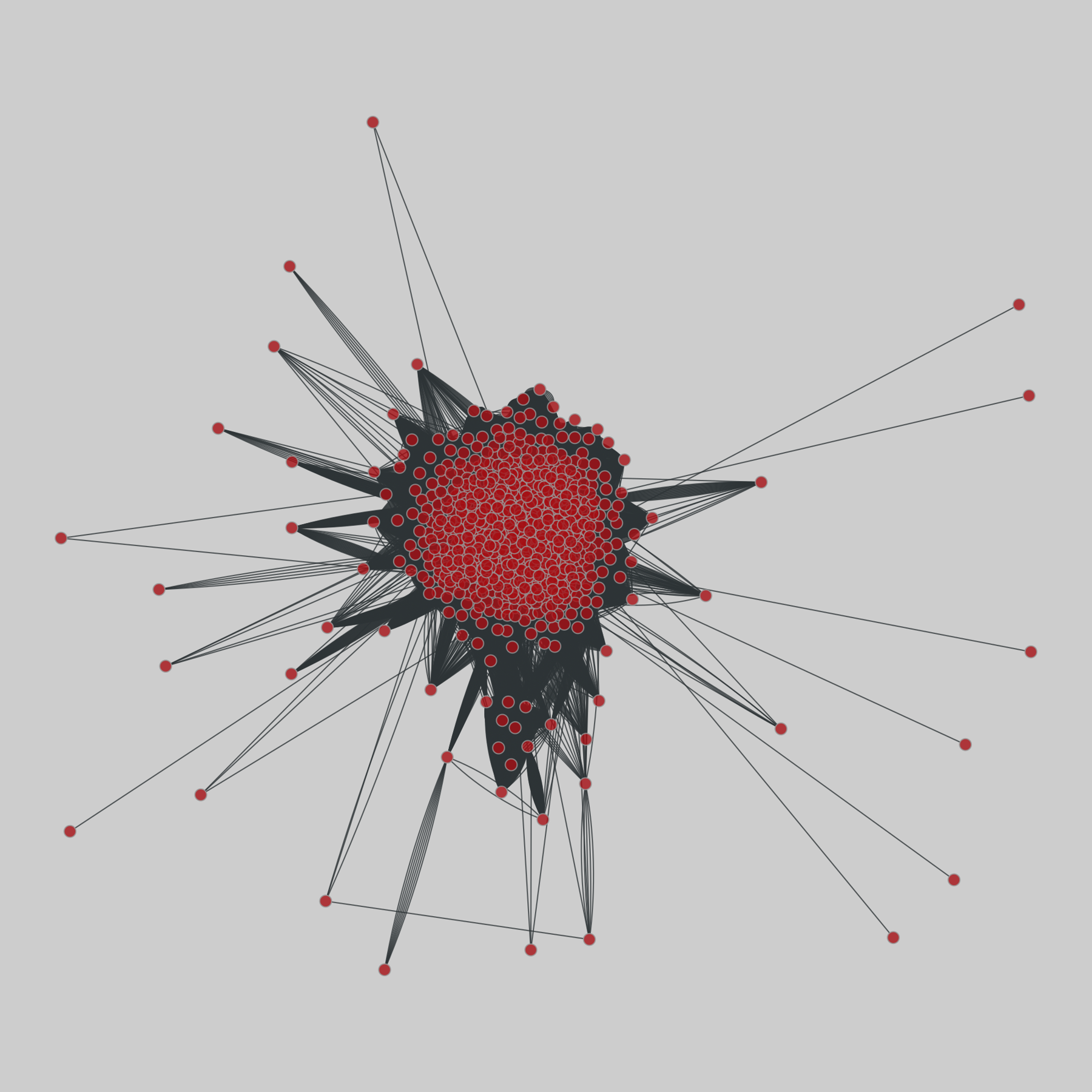
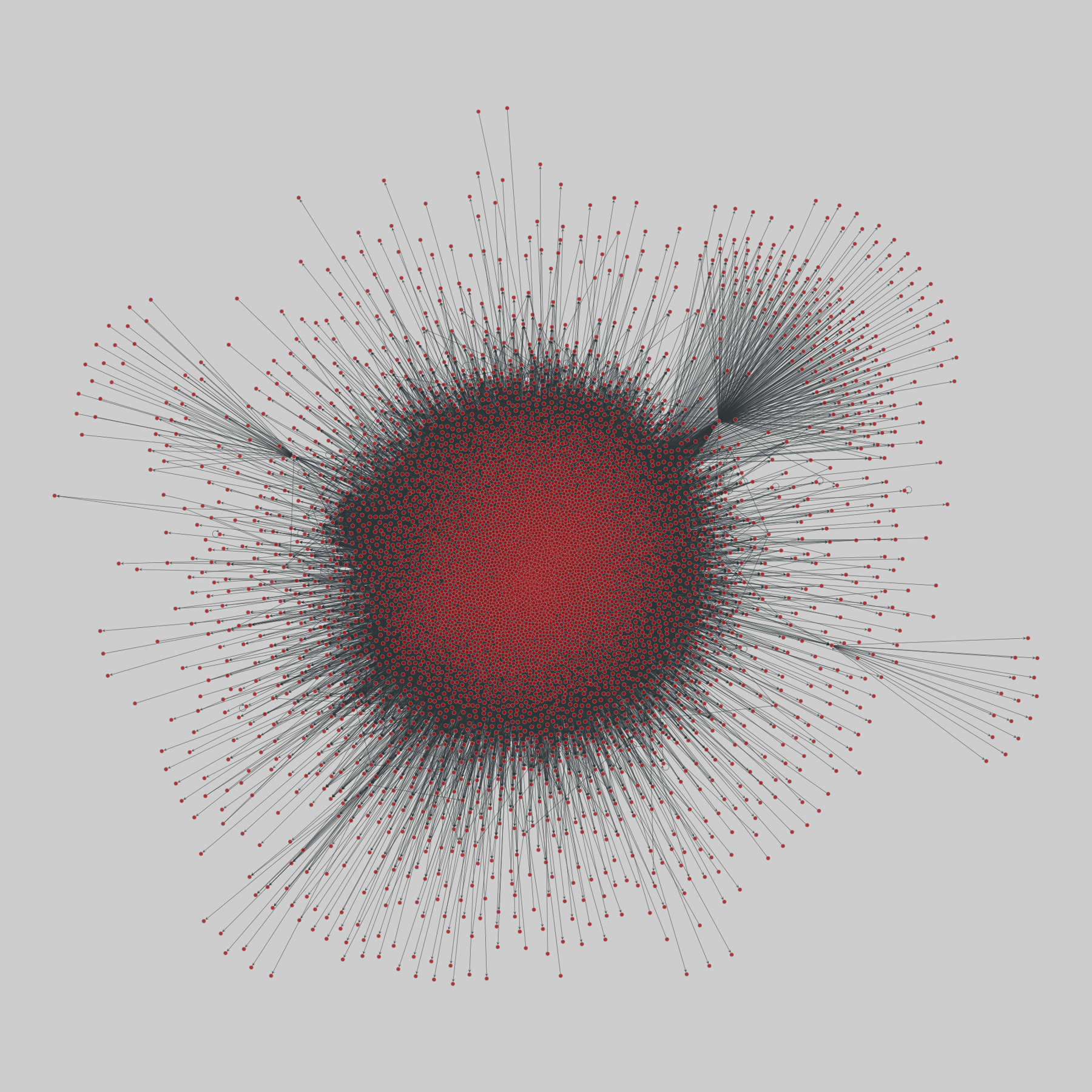
@netzschleuder@social.skewed.degenetic_multiplex: Multiplex genetic interactions (2014)
Multiplex networks representing different types of genetic interactions, for different organisms. Layers represent (i) physical, (ii) association, (iii) co-localization, (iv) direct, and (v) suppressive, (vi) additive or synthetic genetic interaction. Edge direction (i,j) indicates gene i interacting with gene j.
This network has 6570 nodes and 282755 edges.
Tags: Biological, Gene regulation, Protein interactions, Unw…
 @davidaugust@mastodon.online @Techmeme@techhub.social @cdarwin@c.im
Federal agents across the United States were told in recent days that the FBI will no longer investigate physical confrontations involving Immigration and Customs Enforcement officers.
The guidance was shared with FBI managers on Thursday, with some ICE agents notified by their FBI counterparts the same day.
The Justice Department and the Department of Homeland Security issued a joint statement denying that any change had taken place.
They insisted the relationship between …
@cdarwin@c.imBehind the public’s mounting worries is a growing sense that Trump isn’t mentally all there.
Physical and mental health aren’t easily separated, especially as one reaches 80. I often can’t remember where I put my wallet and keys or why I’ve entered a room. I also have less patience than I used to have. (I’m less tolerant of long lines, automated phone menus and Republicans.)
But if Trump can’t remember where he put, say, a top-secret memo or why he entered the Situation Room, or …
@Techmeme@techhub.socialNvidia researchers unveil ENPIRE, an agent harness framework that develops robotic self-improvement strategies for physical tasks with minimal human supervision (Jeremy Hsu/Ars Technica)
https://arstechnica.com/ai/2026/06/ai-coding-agents-can-auton…
@Techmeme@techhub.socialXDOF, which is building data pipelines, collection tools, and annotation systems for robot training data, emerges from stealth with $70M (Tim Fernholz/TechCrunch)
https://techcrunch.com/2026/06/17/colle…
@Techmeme@techhub.socialNvidia unveils Cosmos 3, an open physical AI foundation model, to help robots and autonomous cars better understand the real world with limited training data (Ina Fried/Axios)
https://www.axios.com/2026/06/01/nvidia-ai-push-cosmos-3-world-model
@Techmeme@techhub.socialSony says all new PlayStation games from both first- and third-party developers will be sold in digital formats from January 2028, ending physical game discs (Stephen Totilo/Game File)
https://www.gamefile.news/p/sony-drops-playstation-discs-2028-ps3-vit…
@Techmeme@techhub.socialLG shares jumped 300% this year, after largely sitting out South Korea's chip-rally in 2025, as LG seeks to expand into physical AI businesses such as robots (Sangmi Cha/Bloomberg)
https://www.bloomberg.com/news/articles/20
@Techmeme@techhub.socialA look back at the 2007 heist of a Verizon data center in London, highlighting the role of physical data security as cyber defenses grow more sophisticated (Nathaniel Rich/New York Times)
https://www.nytimes.com/2026/07/12/magazin
@Techmeme@techhub.socialJeff Bezos' Prometheus, which is building AI models for physical tasks, raised a $12B Series B at a $41B valuation, following a $6.2B Series A (Dan Primack/Axios)
https://www.axios.com/2026/06/11/prometheus-bezos-industrial-ai